Endovascular Therapy for Chronic Mesenteric Ischemia


Endovascular Therapy for Chronic Mesenteric Ischemia


Gustavo S. Oderich


Chronic mesenteric ischemia is caused most often by ostial atherosclerotic disease. The typical patient is female, with median age of 65 years old. Most studies quote a 3:1 or 4:1 female-to-male ratio, with age ranging from 40 to 90 years old.


The term intestinal angina has been coined to describe the chronic abdominal pain that occurs within a few minutes to 30 minutes after meals. Patients might describe intolerance to certain types of food, and consequently they alter their eating habits to avoid foods that precipitate symptoms. Unintentional weight loss progresses to malnutrition and cachexia. At times, symptoms are less specific, with vague abdominal pain, nausea, vomiting, or change in bowel habits. Most patients develop the classic symptoms of abdominal pain, weight loss, and food fear once at least two of the three intestinal arteries are severely narrowed or occluded. More than 70% of patients have hypertension, hyperlipidemia, and prior smoking history. Consequently, other manifestations of atherosclerosis are commonplace, including coronary, cerebrovascular, and peripheral arterial disease.


Endovascular treatment has emerged as an alternative to bypass in the elderly or higher-risk patient, but it has become the first choice of treatment in most patients with suitable lesions. The enthusiasm for endovascular techniques is explained by several factors, including high mortality rates (8%–15%) following bypass procedures, the limited experience of vascular surgeons in performing these reconstructions, and the excellent early results of endovascular therapy.


Diagnosis


The diagnosis of chronic mesenteric ischemia is suggested by the clinical history and is confirmed by imaging of the mesenteric arteries. Duplex ultrasound is an excellent screening study. The most used criterion is a peak systolic velocity (PSV) greater than 275 cm/sec for the superior mesenteric artery (SMA) and greater than 200 cm/sec for the celiac artery. Cross-sectional imaging by computed tomography angiography (CTA) or magnetic resonance angiography (MRA) is obtained to identify the extent of mesenteric artery disease and exclude other possible causes of abdominal pain and weight loss.


CTA is very useful to plan the intervention. Key factors that affect choice of approach are the angle of origin of the mesenteric vessels, the amount of calcium and thrombus, and the presence of important collaterals or side branches in proximity to the lesion, such as a replaced hepatic artery arising from the proximal SMA. Conventional angiography remains the gold standard, but it is rarely needed to confirm the diagnosis, except in a minority of patients with questionable disease by CTA or MRA. In most patients, diagnostic angiography is obtained in conjunction with the endovascular procedure. Overall, critical stenosis or occlusion of the SMA is present in more than 90% of patients, of the celiac artery in more than 80%, and of both arteries in more than 80%.


Treatment


Selection


Mesenteric revascularization is indicated in patients with symptoms of chronic mesenteric ischemia and is undertaken rarely as a prophylactic measure in selected patients with asymptomatic three-vessel involvement.


Mesenteric angioplasty and stenting is currently the first choice of treatment in most patients with suitable lesions, independent of their clinical risk. Although there are no randomized comparisons between different types of endovascular techniques, most experts agree that primary stenting is indicated because restenosis is common with angioplasty alone.


The SMA is the primary target for angioplasty and stenting. Technical difficulty is increased by severe calcification, occlusions, long lesions, small vessel diameter, and multiple tandem lesions. Although these anatomic features do not contraindicate the use of stents, one can expect higher rates of local complications, including distal embolization, dissection, and restenosis. Therefore, in the low-risk patient, open surgical bypass should be considered. Other options are celiac stenting or a retrograde hybrid SMA recanalization. Open bypass has been increasingly used in patients who have failed multiple endovascular interventions or who have more extensive disease, with long occlusions or other nonatherosclerotic lesions.


Techniques


Diagnostic Angiography


Diagnostic angiography is usually performed using transfemoral approach with a 5-Fr sheath and 5-Fr diagnostic flush catheter over a 0.035-inch guidewire system. Modest intravenous heparinization (40 units/kg) is recommended before selective catheterization of the mesenteric arteries. The use of a low-osmolar contrast agent, such as iodixanol (Visipaque), minimizes abdominal discomfort during selective injections. A complete study includes abdominal aortogram with anteroposterior and lateral views to define the location, severity, and extent of visceral artery involvement and to identify concomitant lesions in the aorta, renal, or iliac arteries.


The optimal projection to display the proximal celiac artery and SMA is a lateral view, and for the origin of the inferior mesenteric artery (IMA) it is a 15-degree right lateral oblique view. Selective angiography is necessary to confirm the severity of disease and to identify tandem lesions and collateral patterns. A 5-Fr SOS (femoral approach) or MPA catheter (brachial approach) is used. In patients with questionable lesions, pressure gradients can be measured using pressure wire, pull-back, or simultaneous pressure-measurement technique.


Angioplasty and Stenting


The author’s preference is to use a brachial artery approach. This offers excellent support with a small profile system and precise stent deployment in patients with an acute SMA angle. Percutaneous access is established with a micropuncture set using ultrasound guidance. Full systemic heparinization (80 mg/kg) is achieved before catheter manipulations to produce an activated clotting time of more than 250 seconds. A 6-Fr 90-cm Shuttle Select sheath (Cook Medical, Bloomington, IN) is positioned in the descending thoracic aorta above the celiac artery origin. A 5-Fr MPA catheter is ideal for selective catheterization of the mesenteric arteries. In patients with classic symptoms and suitable lesions by CTA, abdominal aortogram is not needed and the intervention is directed to the target vessel. The initial selective angiography should demonstrate the severity of the stenosis and document the distal branches for comparison with postintervention views.


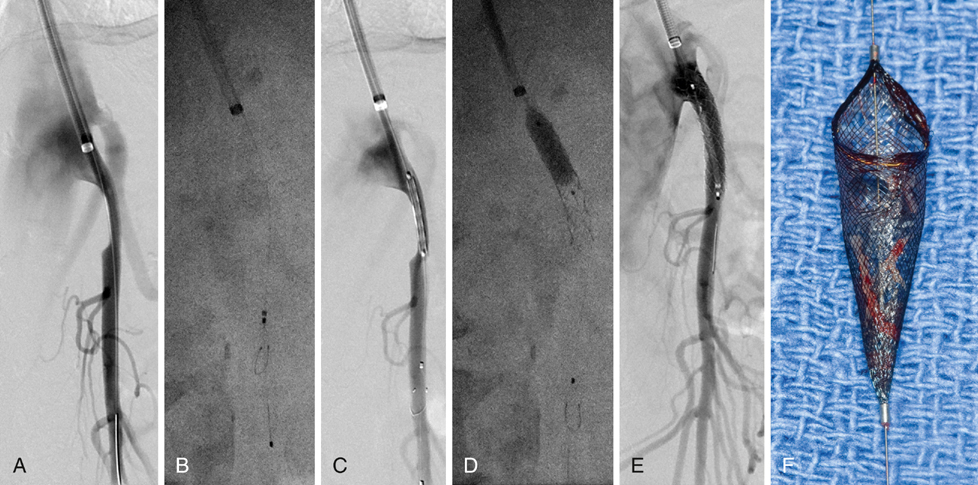
The target SMA lesion is initially crossed using a 0.035-inch soft Glidewire, which is exchanged for the interventional wire of choice after confirmation of true lumen access (Figure 1). The author’s preference is to use a small-profile (0.014- or 0.018-inch) stiff guidewire for most interventions. It is important to visualize the tip of the guidewire, which should be positioned within the main trunk of the SMA rather than within smaller side branches to avoid an inadvertent perforation or dissection. The author’s preference is to use embolic protection (e.g., 320-cm Spider RX filter wire) in patients with occlusions, long lesions (>30 mm), severe calcification, thrombus, and acute or subacute symptoms. If a longer 0.035-inch stent is used, the 0.014-inch filter wire is combined with a 0.018-inch buddy wire, and the stent is introduced via both wires for better support and to facilitate subsequent retrieval of the embolic protection device.


Stay updated, free articles. Join our Telegram channel

Aug 25, 2016 | Posted by in CARDIOLOGY | Comments Off on Endovascular Therapy for Chronic Mesenteric Ischemia

Full access? Get Clinical Tree

Get Clinical Tree app for offline access