Endovascular Radiofrequency Treatment of Varicose Veins


Endovascular Radiofrequency Treatment of Varicose Veins


Steve Elias


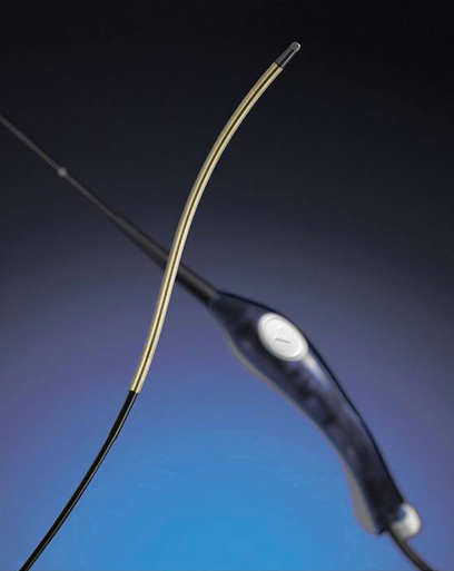
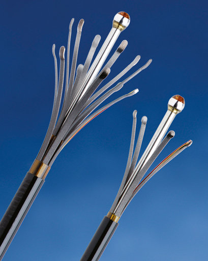
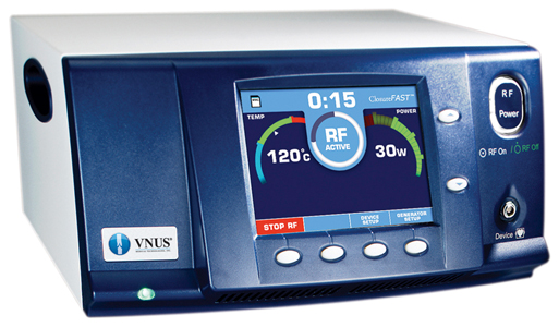
Endovenous radiofrequency (RF) ablation is an effective therapy of varicose veins affecting the greater saphenous vein (GSV), the small saphenous vein (SSV), the anterior accessory greater saphenous vein (AAGSV), the posterior accessory greater saphenous vein (PAGSV), and straight venous segments. The most current therapy is RF-controlled segmental thermal ablation (ClosureFast, Covidien, Mansfield, MA). This catheter fits through a 7-Fr introducer sheath (Figure 1). Venous treatment is segmental, rather than using a continuous catheter pullback rate, resulting in a more standardized delivery of energy. The older RF catheter (Figure 2) used a continuous pullback. The more variables one can eliminate with any technique, the better for physician and patient. A temperature of 120°C is attained at the 7-cm heating segment at the catheter tip. This equates to approximately 70 joules per centimeter of vein treated, which is in the same range delivered with laser ablation. The generator (Figure 3) monitors and delivers this energy automatically by modulating electrical power and impedance.


Thus, for current RF technology, pullback is eliminated and energy delivered is standardized. The only variables left are access and positioning. This is a significant advancement over older RF technology and current laser and mechanochemical techniques. The procedure is more efficacious, and procedure time is shortened relative to previous RF treatments. All forms of endovenous thermal ablation involve four steps: access, positioning, tumescence, and treatment.


Procedure


Access


Access for ClosureFast is no different than for any other endovenous technique: percutaneous and ultrasound guided. The vein is accessed at the lowest point of reflux, whether that be at mid thigh or lower calf. Traditionally, the GSV below the knee was not treated for fear of injury to the saphenous nerve; others have been reluctant to treat the SSV as well, with concern for sural nerve or tibial nerve injury. However, as practitioners have become more facile with ultrasound visualization of these nerves and our ability to appropriately place tumescent anesthesia to protect these nerves, treatment to lower venous segments is now possible with minimal nerve injury. Final results including improvement in quality of life measures (Venous Clinical Severity Score) have been reported. For all these reasons, access is recommended at the lowest point of reflux.


The author prefers to use a micropuncture set that includes a 21-gauge needle, 0.018-inch guidewire, and a 4-Fr or 5-Fr cannula. This set adds a small additional cost to the procedure, but the advantage of a smaller needle and a gentler access outweighs this cost. If one cannot access, one cannot treat. The author also uses a 5-mL slip-tip syringe attached to the needle during cannulation. Gentle continuous aspiration is applied as the needle is being placed into the vein. This technique allows immediate flashback once the needle is intraluminal, thus avoiding unnecessary trauma to the vein, which can induce spasm. The traditional arterial Seldinger technique of through-and-through puncture and then pullback into the lumen waiting for blood return is not as efficacious for venous cannulation, especially with smaller veins. Aggressive spearing or harpooning of the vein should be avoided to minimize spasm. Once access is attained with the micropuncture set, a 7-Fr sheath is used.


This access technique is the author’s personal choice. Other experienced physicians use larger needles without any issue. In addition, if more than one vein is to be treated, wire and/or sheath should be successfully placed into all veins before any treatment. If one fully treats the first vein and then attempts access of a second vein, the second vein might already be in spasm and hard or impossible to access.


Positioning


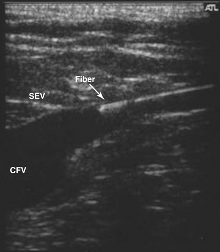
As with all thermal endovenous ablation techniques, the RF catheter is positioned 2 cm from the saphenofemoral junction (SFJ) (Figure 4) or at the fascial curve near the saphenopopliteal junction (SPJ) (Figure 5). In the great majority of cases (>95%) the catheter can be positioned without any adjunctive steps such as guidewire placement, second access site, or ultrasound assistance. In cases of saphenous tortuosity, an attempt can be made to straighten this segment by changing leg position or by using the ultrasound probe with gentle compression to straighten the segment. When doing this, a significant amount of pressure or manipulation should not be applied because it can lead to spasm. Once again, gentle, slow, deliberate movements are best.


Stay updated, free articles. Join our Telegram channel

Aug 25, 2016 | Posted by in CARDIOLOGY | Comments Off on Endovascular Radiofrequency Treatment of Varicose Veins

Full access? Get Clinical Tree

Get Clinical Tree app for offline access