Endobronchial and Endoesophageal Ultrasound Techniques

Chapter 12 Endobronchial and Endoesophageal Ultrasound Techniques


Lung cancer is still the leading cause of cancer deaths worldwide, with an overall 5-year survival rate of 10% to 15%. Mediastinal lymph node sampling in lung cancer is important for adequate staging to determine appropriate treatment, as well as for predicting outcome. Adequate staging of lung cancer also is important in order to improve research into lung cancer, both for accurate comparison of data and for quality control.


Mediastinal lymph node staging can be performed preoperatively by radiologic imaging, endoscopically, or surgically. CT scanning, magnetic resonance imaging (MRI), positron emission tomography (PET), and integrated PET-CT are useful noninvasive imaging techniques for staging of lung cancer; however, they are not sufficiently sensitive or specific to determine mediastinal lymph node involvement. CT scanning usually is the initial method for staging of mediastinal nodes. Only lymph nodes with a short-axis diameter greater than 1 cm (with or without positive findings on PET-CT or other PET study) usually are considered to be suspicious for malignant involvement by radiologic criteria set forth in national and international guidelines. Nevertheless, in view of the high false-positive rate for CT and PET-CT, neither of which provides a tissue diagnosis, it is important to obtain lymph node tissue to determine operability.


Mediastinoscopy with nodal biopsy has been the “gold standard” for mediastinal staging for many years and has a sensitivity of 90% to 95% for detection of metastases in this region. Only certain lymph node stations—2, 4, and anterior 7—are accessible by this approach, in which access to the posterior and inferior mediastinum is limited, typically necessitating extending the procedure to cervical mediastinoscopy or thoracoscopy. However, it is essentially a surgical approach requiring general anesthesia and occasionally hospitalization. Endoscopic techniques provide a minimally invasive alternative to surgical staging. Accordingly, the past few years have seen the development of a number of less invasive staging modalities, including endobronchial ultrasound (EBUS) and endoesophageal ultrasound (EUS) techniques.


Transbronchial Needle Aspiration


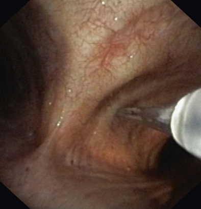
Merely a curiosity at its inception, flexible bronchoscopy has emerged as an essential diagnostic and therapeutic modality in the management of a variety of lung diseases. The addition of transbronchial needle aspiration (TBNA) not only improved bronchoscopy’s diagnostic yield but further extended the role of this modality in the evaluation of mediastinal disease, and in the diagnosis and staging of bronchogenic carcinoma. The first description of sampling mediastinal lymph nodes through the tracheal carina using a rigid bronchoscope was by Schieppati. In 1978, Wang and associates demonstrated that it was feasible to sample paratracheal nodes using TBNA. Subsequent publications highlighted the use of the technique in the diagnosis of endobronchial (Figure 12-1) and peripheral lesions and the ability of TBNA to provide a diagnosis even in the absence of endobronchial disease.


The diagnostic yield of TBNA in the assessment of hilar-mediastinal lymph node involvement in lung cancer varies greatly in published series, with reported rates ranging from 15% to 85%. Recently, a metaanalysis assessing TBNA for mediastinal staging in non–small cell lung cancer demonstrated that TBNA is highly specific for the identification of mediastinal metastases, with sensitivity depending heavily on the study population under investigation. In studies that included patient populations with a prevalence of mediastinal metastases of 34%, sensitivity was only 39%, whereas in a population with a prevalence of 81%, sensitivity for detection of metastases was 78%.


Nevertheless, even after more than 50 years since the advent of TBNA, the technique is still underused. The main reasons for the limited use of TBNA have been lack of needle monitoring, difficulties in performing the procedure, and a belief, despite good evidence to the contrary, that TBNA is not useful.


Endobronchial Ultrasound Technique


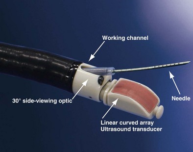
The integration of ultrasound technology and flexible fiberoptic bronchoscopy enables imaging of lymph nodes, lesions, and vessels located beyond the tracheobronchial mucosa. Developed in 2002, the EBUS bronchoscope looks similar to a normal bronchovideoscope (Figure 12-2) but is 6.9 mm wide and has a 2-mm instrument channel and a 30-degree side viewing optic. Furthermore, a curved linear array ultrasound transducer sits on the distal end and can be used either with direct contact to the mucosal surface or with an inflatable balloon that can be attached at the tip. This setup produces a conventional endoscopic picture side by side with the ultrasound view. Ultrasound scanning is performed at a frequency of 7.5 to 12 MHz, with tissue penetration of 20 to 50 mm. An ultrasound processor generates the ultrasound image.


EBUS allows the bronchoscopist to visualize airway structures as well as surrounding processes. It is useful for staging advanced cancer, especially as it relates to intramural or nodal spread. EBUS can identify N1, N2, and N3 nodes without the need for surgical intervention and can hence decrease the need for expensive surgery.


Procedure


The actual TBNA is performed using direct transducer contact with the wall of the trachea or bronchus. When a lesion is outlined, a 21 or 22 gauge needle can be advanced through the working channel, and lymph nodes can be punctured under real-time ultrasound visualization. The needle is encased in an internal sheath in order to avoid contamination during biopsy. At the same time, color Doppler can be used to identify surrounding vascular structures. Once the target lymph node or mass has been clearly identified with EBUS, the needle is inserted under real-time ultrasound guidance and then placed within the lesion (Figures 12-3 and 12-4). Suction is applied with a syringe, and the needle is moved back and forth to achieve multiple punctures. The stylet of the needle is left in place on the first puncture to minimize bronchial cell contamination; once the needle tip is inside the target tissue, the stylet is removed. We stab the target 10 to 15 times without suction and apply suction only for the last two or three stabbing motions. Before retraction of the needle into the needle sheath, suction must be removed to minimize sample loss into the syringe. The specimen is then air-flushed onto a slide, and the needle is flushed with heparin-saline solution to avoid clotting; the same procedure is repeated three times at every lymph node station.
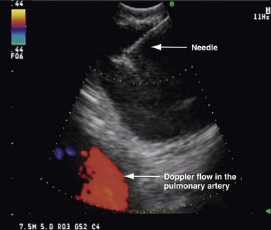
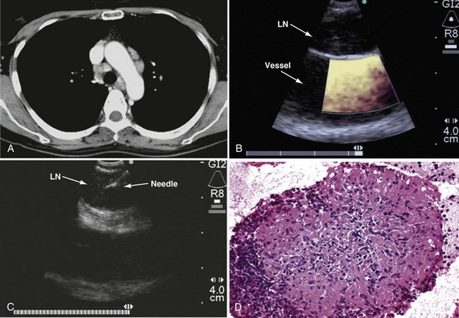


Stay updated, free articles. Join our Telegram channel

Jun 12, 2016 | Posted by in RESPIRATORY | Comments Off on Endobronchial and Endoesophageal Ultrasound Techniques

Full access? Get Clinical Tree

Get Clinical Tree app for offline access