EMBRYOLOGY OF THE HEART
Introduction
Cardiovascular development begins early in fetal life around the fourth week of gestation and continues through the tenth week. During this time, the heart begins to form, starting from the cardiogenic fields and ending up as a 4-chambered structure. During these weeks, initially a tube, the heart elongates and loops, has septation, loses its symmetry, develops 2 separate outflow tracts connected to the 2 great arteries, and forms venous structures, aortic arches, and specialized tissue for conduction. To be able to understand the cardiac malformations that develop in the fetus, it is imperative to have knowledge of the developing heart from the first day it begins its formation to completion by the tenth week after conception. This chapter summarizes the steps in cardiac formation, which will then help to understand the background for the development of congenital cardiac defects.
Early Cardiogenesis
The vascular system, which appears in the embryo in the middle of the third week, is the first major organ system to develop and become fully functional in the embryo. Its nutrition is supplied by diffusion until vasculature develops. Circulation must replace diffusion for the embryo to develop beyond the germ layer. The cardiogenic precursors that lie in the epiblast migrate through the primitive streak and form the mesodermal cardiogenic fields (Figure 3.1).
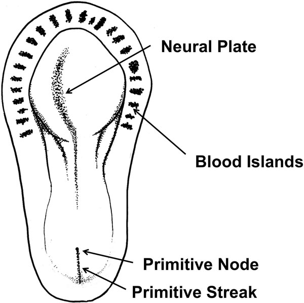
Figure 3.1. Presomite embryo. Cardiac progenitor cells in the epiblast layer migrate through the streak to their destination. They are induced to form the myoblasts. The blood islands are in front of the neural crest and the sides of the embryo.
The cells that will form the cardinal segments of the heart and the outflow tracts migrate first and reside in the splanchinic layer of the lateral plate mesoderm, where they are induced by the pharyngeal endoderm to form cardiac myoblasts. The cells forming the caudal parts of the heart such as the right ventricle (RV), left ventricle (LV), and the sinus venosus migrate in a sequential order.1 The cells in the primitive streak are initially organized rostrocaudally and change to mediolateral organization after migration. Mesoderm also houses the blood islands that form the blood cells and vessels with a process called vasculogenesis. The cardiogenic field, which is a horseshoe-shaped tube lined with the endothelium, is surrounded by myoblasts (Figure 3.2). Other blood islands that appear bilateral, parallel, and close to the embryonic shield acquire a lumen and form a pair of longitudinal vessels, the dorsal aorta which then connects to the horseshoe-shaped plexus to form the heart tube.1
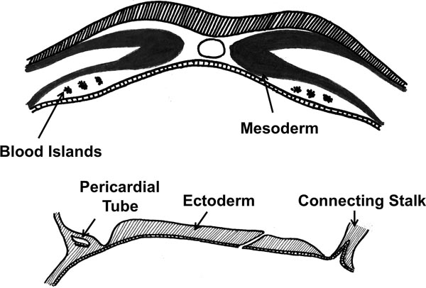
Figure 3.2. Vasculogenesis. The blood islands in the mesoderm form the blood cells and vessels.
Formation of the Heart Tube
Bilateral endocardial heart tubes form in the third week of development (approximately day 18). Lateral folding of the embryo brings the heart tubes into the ventral midline, allowing them to fuse to form a single primordial heart tube. Fusion of the heart tubes begins cranially and extends caudally except at the very end. With the fusion, the heart becomes a continuous tube that receives venous drainage from the caudal pole and pumps blood out of the first aortic arch to the dorsal aorta at its cranial pole (Figure 3.3).
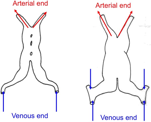
Figure 3.3. Formation of the heart tube. Bilateral heart tubes start to fuse from cranial to caudal and become a continuous tube with venous and arterial ends.
Around day 22, the fused heart tube protrudes into the pericardial cavity and is suspended at its cranial and caudal poles with the help of blood vessels. It consists of three different layers—the myocardium, lined by the endocardium internally and the epicardium externally.2 Myocardium forms from the splanchnic mesoderm. During looping, additional myocardial cells are added to the outflow tract. The endocardium forms from the endothelium of the heart tube while the epicardium develops from the mesothelial cells that arise from the sinus venosus. It is also at this time that the heart begins to beat. 3
Once fusion is complete and before looping begins, some constrictions and dilations appear in the heart tube to form certain initial divisions of the primitive heart. From cranial to caudal, these sections are truncus arteriosus (TA), bulbus cordis, primordial ventricle, primordial atrium, and sinus venous (Figure 3.4).
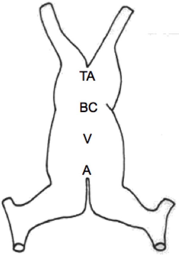
Figure 3.4. Sections of heart tube just before looping. After the completion of fusion of the heart tubes, various sections may be designated as TA, truncusarteriosus;BC, bulbuscordis;V, primordial ventricle; and A, primordial atrium.
The atrial portion of the cardiac tube becomes the common atrium; bulbus cordis is divided into 3 parts. The proximal portion forms the trabeculated RV; conus cordis, which is the mid-portion, forms the outflow tracts, and the truncus arteriosus forms the arterial roots, proximal aorta and the pulmonary artery (PA) as its distal portion. 1
Looping
Cardiac looping starts in the fourth week and is completed during the fifth week of development. Since there is not enough space to accommodate the lengthening of the straight heart tube, it starts to bend as it elongates. The simultaneous growth of the bulbus cordis and the primitive ventricle forces the heart to bend ventrally and rotate to the right.
The cephalic portion bends ventrally, caudally, and to the right (D-loop) while the caudal (atrial) portion moves dorsocranially and to the left1 (Figures 3.5 and 3.6). If the heart tube loops to the left (L-loop), the morphologic RV moves to the left side, causing ventricular inversion which is associated with L-transposition or corrected transposition.3
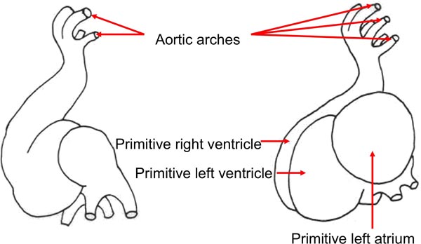
Figure 3.5. Looping of the heart tube (viewed from side).
Stay updated, free articles. Join our Telegram channel

Full access? Get Clinical Tree


