Electrocardiographic voltage has been used to determine the presence of left ventricular hypertrophy for about 70 years. Varying electrocardiographic criteria have been applied. We have found total 12-lead QRS voltage to be most useful in this regard. We measured total 12-lead QRS voltage in 24 patients in whom an ascending aortic aneurysm was resected and histologic study of its wall was classic of syphilitic aortitis. In these 24 patients total 12-lead QRS voltage ranged from 57 to 161 mm, averaging 120 ± 32 in the 11 men and 106 ± 24 mm in the 13 women. If normal 12-lead QRS voltage in adults is considered to be >175 mm not a single one of the 24 patients had normal voltage. Indeed, most were in the low normal area. Thus, this study provides some evidence via this indirect means that the heart itself is infrequently involved by syphilitic aortitis which produces an ascending aortic aneurysm of sufficient size to warrant resection.
During the past 30 years we have compared electrocardiographic total 12-lead QRS voltage to heart weight at necropsy or after cardiac transplantation in 11 different cardiac conditions. These studies were summarized in a recent review, which also demonstrated that total 12-lead QRS voltage was the best electrocardiographic indicator of increased cardiac mass. Our necropsy studies in patients with syphilitic aortitis have indicated that, with few exceptions, the heart is of normal size. The present study examines total 12-lead QRS voltage in 24 patients who underwent operative resection of a thoracic syphilitic aneurysm to determine if any had evidence of increased cardiac mass using this indirect criterion.
Methods
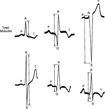
From 2010 through 2014, twenty-four patients have had resection of a syphilitic aneurysm involving the ascending aorta at Baylor University Medical Center at Dallas, and each had an immediate preoperative electrocardiogram available for examination. The QRS voltage (from the peak of the R wave to the nadir of either the Q or the S wave, whichever was deeper [ Figure 1 ]) was measured in each of the 12 leads in each of the 24 patients and certain other clinical features also were collected.
Results
Pertinent findings in each of the 24 patients are summarized in Table 1 . Total 12-lead QRS voltage (measured with normal [10 mm] standardization [10 mm = 1 mV]) in the 11 men (mean age 61 ± 17 years) ranged from 57 to 161 mm (mean 120 ± 32), and in the 13 women (mean age 71 ± 9 years), from 64 to 146 mm (mean 106 ± 24). All but one of the 24 patients had a history of systemic hypertension. Echocardiograms, available for review in 4 of the 24 patients, disclosed left ventricular wall thicknesses ≤1.2 cm in all 4 patients, left ventricular ejection fractions ≥55% in all 4, aortic regurgitation in 4 (all minimal or mild), and a dilated left ventricular cavity in none. Computed tomographic imaging preoperatively, data available in 15 of the 24 patients, showed the maximal diameter of the ascending aorta to range from 4.7 to 8.2 cm (mean 6.5 in 6 men and 5.7 in 9 women). The body mass index (Kg/m 2 ) ranged from 23 to 38 (mean 28 ± 4) in the 11 men, and from 15 to 47 (mean 28 ± 9) in the 13 women. Data from coronary angiography, available in 18 patients, disclosed narrowing of 1 or more major coronary arteries in 6 (33%) patients and 4 of the 6 underwent coronary artery bypass grafting. Serum total cholesterol levels (15 patients) ranged from 79 to 225 mg/dl (mean 163) and was >200 mg/dl in only 3 patients; low-density lipoprotein cholesterol ranged from 39 to 164 mg/dl (mean 100) and was >100 mg/dl in 6 of the 15 patients.
| # | Age (years) | RACE | BMI (Kg/m2) | QRS Amplitude in Each Electrocardiographic lead | TC (mg/dl) | LDL (mg/dl) | Coronary Angiography Done | No. of CA Narrrowed | CT Asc Aorta An Diameter (cm) | Cardiac Catheterization | AVR | ||||||||||||||||
|---|---|---|---|---|---|---|---|---|---|---|---|---|---|---|---|---|---|---|---|---|---|---|---|---|---|---|---|
| I | II | III | aVR | aVL | aVF | V1 | V2 | V3 | V4 | V5 | V6 | QRSTOTAL | BBB | LV (s/d) (mmHg) | SA (s/d) (mmHg) | EF (%) | CI (L/min/m 2 ) | ||||||||||
| MEN | |||||||||||||||||||||||||||
| 1 | 33 | W | 24.7 | 11 | 9 | 5 | 9 | 9 | 5 | 12 | 17 | 12 | 22 | 23 | 17 | 151 | 0 | 184 | 96 | + | 0 | 133/26 | 133/73 | 60 | 3.1 | + | |
| 2 | 45 | B | 23 | 3 | 12 | 10 | 6 | 5 | 11 | 11 | 10 | 21 | 25 | 17 | 11 | 142 | 0 | – | – | 0 | – | 8.2 | – | 180/100 (I) | 55 (E) | – | 0 |
| 3 | 48 | B | 27.9 | 8 | 10 | 6 | 9 | 3 | 9 | 13 | 16 | 9 | 21 | 21 | 15 | 140 | 0 | 218 | 147 | 0 | – | 6.2 | – | 121/77 (I) | 60 (E) | – | 0 |
| 4 | 51 | W | 28.1 | 9 | 5 | 5 | 7 | 7 | 4 | 12 | 11 | 20 | 34 | 29 | 18 | 161 | 0 | 151 | 97 | + | 0 | 7.4 | 144/22 | 131/60 | 35 | 2.4 | 0 |
| 5 | 59 | A | 28.4 | 7 | 7 | 5 | 6 | 5 | 4 | 7 | 12 | 12 | 12 | 14 | 16 | 152 | 0 | 186 | 118 | 0 | – | 4.7 | – | 120/80 (I) | 74 (E) | – | 0 |
| 6 | 59 | B | 34.4 | 5 | 7 | 3 | 5 | 2 | 4 | 2 | 6 | 18 | 25 | 23 | 18 | 118 | 0 | – | – | 0 | – | AD | – | 111/65 (I) | 65 (E) | – | + |
| 7 | 60 | W | 28.9 | 8 | 2 | 7 | 5 | 8 | 4 | 5 | 15 | 17 | 18 | 11 | 9 | 109 | 0 | 225 | 156 | + | 0 | – | 145/23 | 14/85(I) | 63 | 1.6 | 0 |
| 8 | 70 | W | 37.7 | 9 | 3 | 8 | 6 | 9 | 3 | 7 | 9 | 11 | 12 | 12 | 13 | 102 | 0 | 136 | 88 | + | 3(CABG) ∗ | 6.7 | 173/33 | 170/33 | 45 | – | 0 |
| 9 | 80 | W | 28 | 6 | 4 | 6 | 3 | 6 | 5 | 7 | 6 | 15 | 21 | 18 | 11 | 108 | +R | 108 | 49 | + | 1 (CABG) | 5.7 | 129/9 | 130/59 | 55 | 3.4 | 0 |
| 10 | 83 | W | 24.4 | 6 | 19 | 5 | 5 | 6 | 2 | 5 | 5 | 10 | 11 | 3 | 9 | 86 | 0 | 128 | 81 | + | 2 (CABG) ∗ | – | 133/10 | 132/59 | 35 | 3.2 | + |
| 11 | 84 | W | 26.5 | 5 | 5 | 4 | 4 | 4 | 3 | 6 | 4 | 4 | 5 | 7 | 6 | 57 | 0 | 126 | 79 | + | 1 (CABG) | – | 121/19 | 103/63 | 45 | – | + |
| 33-84 | 23-37.7 | 57-161 | |||||||||||||||||||||||||
| (61±17) | (28.4±4.3) | (120±32) | |||||||||||||||||||||||||
| WOMEN | |||||||||||||||||||||||||||
| 1 | 58 | B | 31.7 | 4 | 7 | 9 | 5 | 6 | 8 | 4 | 11 | 10 | 14 | 15 | 10 | 103 | 0 | 184 | 110 | + | 0 | 5.1 | 126/3 | 125/80 | 60 | – | 0 |
| 2 | 59 | W | 28.9 | 19 | 15 | 4 | 14 | 9 | 6 | 11 | 9 | 13 | 16 | 16 | 14 | 146 | 0 | 190 | 110 | + | 0 | 6.6 | – | 156/83 | 60 (E) | – | + |
| 3 | 62 | W | 45.6 | 8 | 6 | 4 | 7 | 5 | 4 | 14 | 17 | 25 | 9 | 11 | 12 | 122 | 0 | 199 | 141 | + | 0 | 5.1 | 152/26 | 153/45 | 50 | 2.7 | + |
| 4 | 65 | W | 46.5 | 6 | 4 | 3 | 5 | 4 | 2 | 3 | 12 | 8 | 11 | 12 | 8 | 78 | 0 | 129 | 65 | + | 0 | 6.4 | 114/16 | 117/67 | 65 (E) | – | 0 |
| 5 | 65 | W | 28.9 | 7 | 4 | 6 | 6 | 7 | 3 | 7 | 12 | 17 | 19 | 20 | 14 | 122 | 0 | 129 | 65 | 0 | – | 6 | 153/13 | 154/83 | 70 | – | 0 |
| 6 | 67 | W | 26.2 | 12 | 9 | 8 | 10 | 9 | 6 | 12 | 16 | 13 | 11 | 10 | 9 | 125 | 0 | 86 | 47 | + | 0 | – | 124/4 | 125/83 | 60 | 2.9 | 0 |
| 7 | 69 | W | 15.8 | 2 | 10 | 8 | 6 | 4 | 10 | 4 | 4 | 5 | 10 | 14 | 23 | 100 | 0 | 158 | 84 | + | 2 | 5.6 | 101/3 | 100/60 | 55 | – | 0 |
| 8 | 70 | W | 21.6 | 5 | 6 | 4 | 6 | 3 | 4 | 4 | 10 | 15 | 18 | 25 | 14 | 114 | 0 | 142 | 69 | + | 0 | 5.4 | – | 156/57 | 63 (E) | – | 0 |
| 9 | 78 | B | 25.1 | 9 | 8 | 8 | 7 | 7 | 6 | 10 | 6 | 9 | 13 | 15 | 12 | 111 | 0 | 167 | 112 | + | 1 (CABG) ∗ | – | – | 134/88 (I) | – | – | + † |
| 10 | 79 | W | 24.8 | 5 | 7 | 4 | 6 | 3 | 5 | 9 | 7 | 5 | 4 | 4 | 11 | 70 | 0 | – | – | + | 3 | 5.2 | – | 143/57 (I) | 60 (E) | – | + |
| 11 | 80 | W | 28.6 | 4 | 11 | 12 | 4 | 6 | 10 | 6 | 9 | 7 | 9 | 10 | 11 | 99 | 0 | – | – | + | 0 | 5.7 | 144/27 | 140/54 | 60 | 3.1 | 0 |
| 12 | 83 | W | 22.3 | 5 | 4 | 3 | 4 | 4 | 3 | 4 | 11 | 3 | 10 | 8 | 5 | 64 | 0 | – | – | + | 0 | – | 124/13 | 123/58 | 60 | 3.7 | 0 |
| 13 | 83 | W | 18.6 | 8 | 6 | 4 | 6 | 5 | 6 | 10 | 10 | 14 | 23 | 17 | 11 | 120 | +R | 79 | 39 | + | 0 | – | 182/18 | 179/54 | 55 | 2.8 | + |
| 58-83 | 15.8-46.5 | 64-146 | |||||||||||||||||||||||||
| (71±9) | (28.0±9.1) | (106±24) | |||||||||||||||||||||||||
Stay updated, free articles. Join our Telegram channel

Full access? Get Clinical Tree


