Electrocardiographic Interpretation
Donald A. Underwood
CODING SHEET
General Features
1.Normal electrocardiogram (ECG)
2.Borderline ECG or normal variant (specify in other section)
3.Incorrect electrode placement
P-Wave Abnormalities
4.Right atrial abnormality
5.Left atrial abnormality
6.Nonspecific atrial abnormality
7.Sinoventricular condition with absent P wave
Supraventricular Rhythms
8.Normal sinus rhythm (without other abnormalities of rhythm or atrioventricular (AV) conduction)
9.Sinus rhythm (in presence of abnormality of rhythm or AV conduction)
10.Sinus arrhythmia
11.Sinus bradycardia
12.Sinus tachycardia
13.Sinus pause or arrest
15.Ectopic atrial or junctional rhythm
16.Wandering atrial pacemaker
17.Atrial premature beats, normally conducted
18.Atrial premature beats, nonconducted
19.Atrial premature beats with aberrant intraventricular conduction
20.Atrial tachycardia (regular, sustained, 1:1 conduction)
21.Atrial tachycardia, repetitive (short paroxysms)
22.Atrial tachycardia, multifocal (chaotic atrial tachycardia)
23.Atrial tachycardia with AV block
24.Atrial flutter
25.Atrial fibrillation
26.Retrograde atrial activation
27.Supraventricular tachycardia, unspecified
Atrioventricular Junctional Rhythms
28.AV junctional premature beats
29.AV junctional escape beats or escape rhythm
30.AV junctional rhythm, accelerated rhythm (nonparoxysmal junctional tachycardia)
31.AV junctional tachycardia
Ventricular Rhythms
32.Ventricular premature beat(s), uniform, fixed coupled
33.Ventricular premature beats, R on T phenomenon
34.Premature ventricular contractions, in pairs
35.Ventricular parasystole
36.Ventricular tachycardia
37.Accelerated idioventricular rhythm
38.Ventricular escape beats or rhythm
39.Ventricular fibrillation
Atrioventricular Conduction Abnormalities
(Also see items 48 to 53)
40.AV block, primary
41.AV block, secondary—Mobitz type I (Wenkenbach)
42.AV block secondary—Mobitz type II
43.AV block, 2;1, 3:1, 4:1
44.AV block, complete
45.AV block, varying
46.Short PR interval (with sinus rhythm and normal QRS duration)
47.Preexcitation (Wolff-Parkinson-White) syndrome(s)
Atrial Ventricular Interactions in Arrhythmias
(Also see items 40 to 47)
48.Fusion beats
49.Reciprocal (echo) beats
50.Ventricular capture beats
51.AV dissociation (without complete AV block)
52.Isorhythmic AV dissociation
53.Ventriculophasic sinus arrhythmia
Abnormalities of QRS Voltage or Axis
54.Low voltage, limb leads only
55.Low voltage, limb and precordial leads
56.Left axis deviation (>30 degrees)
57.Right axis deviation (>+100 degrees)
58.Electrical alternans
Ventricular Hypertrophy
59.Left ventricular hypertrophy by voltage only
60.Left ventricular hypertrophy by left ventricular hypertrophy by voltage and ST-T segment (secondary repolarization changes)
61.Right ventricular hypertrophy
62.Combined ventricular hypertrophy
Intraventricular Conduction Disturbances
63.Right bundle branch block (RBBB), incomplete
64.RBBB, complete
65.Left anterior fascicular block
66.Left posterior fascicular block
67.Left bundle branch block (LBBB), complete with ST-T waves suggestive of acute myocardial injury or infarction
68.LBBB, complete
69.Intraventricular conduction disturbance, nonspecific type
70.Aberrant intraventricular conduction with supraventricular arrhythmia (specify rhythm)
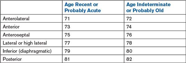
Transmural Myocardial Infarction
(Also see items 88 and 89)

83.Probable ventricular aneurysm
ST-, T-, and U-Wave Changes
84.Subendocardial or subepicardial nontransmural infarction
85.Normal variant, early repolarization
86.Normal variant, juvenile T wave
87.Nonspecific ST- and/or T-wave changes
88.ST- and/or T-wave changes suggesting myocardial ischemia
89.ST- and/or T-wave changes suggesting myocardial injury
90.ST- and/or T-wave changes suggesting acute pericarditis
91.ST-T segment changes secondary to intraventricular conduction distribution or hypertrophy
92.Post extrasystolic T waves
93.Isolated J-point depression
94.Peaked T waves
95.Prolonged QT interval
96.Prominent U waves
Suggested Probable Clinical Disorder
97.Digitalis effect
98.Digitalis toxicity
99.Hyperkalemia
100.Hypokalemia
101.Hypercalcemia
102.Hypocalcemia
103.Atrial septal defect, secundum
104.Atrial septal defect, primum
105.Dextrocardial, mirror image
106.Mitral valve disease
107.Chronic lung disease
108.Acute cor pulmonale including pulmonary embolus
109.Pericardial effusion
110.Acute pericarditis
111.Hypertrophic obstructive cardiomyopathy (idiopathic hypertrophic subaortic stenosis)
112.Hypertrophic obstructive cardiomyopathy; apical
113.Coronary artery disease
114.Central nervous system (CNS) disorder
115.Myxedema
116.Hypothermia
Stay updated, free articles. Join our Telegram channel

Full access? Get Clinical Tree


