A low ratio of n-3 to n-6 polyunsaturated fatty acids has been associated with cardiovascular events. However, the effects of this ratio on coronary atherosclerosis have not been fully examined. The purpose of the present study was to evaluate the correlations between the n-3 to n-6 polyunsaturated fatty acid ratio and coronary atherosclerosis. Coronary atherosclerosis in nonculprit lesions in the percutaneous coronary intervention vessel was evaluated using virtual histology intravascular ultrasound in 101 patients at the time of percutaneous coronary intervention and 8 months after statin therapy. Forty-six patients (46%) showed atheroma progression and the remaining 55 patients (54%) showed atheroma regression at 8-month follow-up. Significant negative correlations were observed between percentage change in plaque volume and change in the eicosapentaenoic acid (EPA)/arachidonic acid (AA) ratio (r = −0.190, p = 0.05), docosahexaenoic acid (DHA)/AA ratio (r = −0.231, p = 0.02), and EPA+DHA/AA ratio (r = −0.240, p = 0.02). Furthermore, percentage change in the fibrous component volume was negatively and significantly correlated with change in the EPA/AA ratio (r = −0.206, p = 0.04) and EPA+DHA/AA ratio (r = −0.217, p = 0.03). Multivariate regression analysis showed that change in the EPA+DHA/AA ratio was a significant predictor of percentage change in plaque volume and fibrous component volume (β = −0.221, p = 0.02, and β = −0.200, p = 0.04, respectively). In conclusion, decreases in serum n-3 to n-6 polyunsaturated fatty acid ratios are associated with progression in coronary atherosclerosis evaluated using virtual histology intravascular ultrasound in statin-treated patients with coronary artery disease.
Intensive lipid-lowering therapy with statins results in regression or stabilization of coronary artery plaques and reduces the risk for coronary events. However, not all patients show regression or stabilization of coronary atherosclerosis after statin therapy. Several studies have reported that consumption of fish and fish oil containing large concentrations of n-3 polyunsaturated fatty acids (PUFAs) is associated with a low risk for cardiovascular disease. Among the n-3 PUFAs, eicosapentaenoic acid (EPA) and docosahexaenoic acid (DHA) play important roles in preventing cardiovascular disease. The Japan EPA Lipid Intervention Study (JELIS), a large randomized control trial, demonstrated that pure EPA administration along with statin therapy decreased the incidence of coronary events by 19%. On the basis of these reports, the residual risk for cardiovascular events after statin therapy can be explained in part by a low ratio of n-3 to n-6 PUFAs. Therefore, we performed a post hoc analysis of the Treatment With Statin on Atheroma Regression Evaluated by Intravascular Ultrasound With Virtual Histology (TRUTH) study to investigate the relation between n-3 to n-6 PUFA ratio and coronary atherosclerosis using serial virtual histology (VH)–intravascular ultrasound (IVUS) measurements before and after statin therapy.
Methods
The present study was a post hoc analysis of the TRUTH study. The TRUTH study was a prospective, open-labeled, randomized, multicenter trial performed at 11 Japanese centers to evaluate the effects of 8-month treatment with pitavastatin versus pravastatin on the composition of coronary artery plaques using VH-IVUS. Briefly, 164 patients with angina pectoris were randomized to either pitavastatin (4 mg/day; intensive lipid lowering) or pravastatin (20 mg/day; moderate lipid lowering ) therapy after successful percutaneous coronary intervention (PCI) under VH-IVUS guidance. Follow-up IVUS was performed after 8 months of statin therapy.
Patients were included in the present study if they fulfilled the following criteria: coronary plaque of a non-PCI site in a culprit vessel evaluated using VH-IVUS at PCI and at 8-month follow-up along with adequate serum volume in frozen samples for various measurements. A total of 101 patients were included in this study. These patients were divided into 2 groups: progressors and regressors. A progressor was defined as a patient with plaque volume at 8-month follow-up minus plaque volume at baseline ≥0. A regressor was defined as a patient with plaque volume at 8-month follow-up minus plaque volume at baseline <0. We compared serum n-3 and n-6 PUFA levels and their ratios in progressors and regressors; moreover, we investigated the relation between these ratios and coronary atherosclerosis.
Details regarding the IVUS procedure have been documented. Briefly, after PCI of the culprit lesion, IVUS was performed for angiographic lesions with <50% luminal narrowing on the distal and proximal sides of the culprit lesion. An IVUS catheter (Eagle Eye Gold; Volcano Corporation, San Diego, California) was used, and a motorized pullback device was used to withdraw the transducer at 0.5 mm/s. During pullback, grayscale IVUS was recorded, and raw radiofrequency data were captured at the top of the R wave using a commercially available IVUS console (IVG3; Volcano Corporation). After 8 months of statin therapy, IVUS was repeated in the same coronary artery using the same type of IVUS catheter used at baseline.
All baseline and follow-up IVUS core laboratory analyses were performed by an independent and experienced investigator (M.T.) in a blinded manner. Before IVUS analysis, baseline and follow-up IVUS images were reviewed side by side on a display, and distal and proximal ends of the target segment were identified on the basis of the presence of reproducible anatomic landmarks such as the side branch, vein, and stent edge. Plaques close to the PCI site (<5 mm) were excluded because mechanical interventions affected atheroma measurements. Manual contour detection of the lumen and external elastic membrane was performed for each frame. Quantitative IVUS grayscale analysis was performed according to the guidelines of the American College of Cardiology and European Society of Cardiology. VH-IVUS data analysis was based on grayscale border contour calculation, and relative and absolute amounts of different coronary artery plaque components were measured using IVUSLab version 2.2 (Volcano Corporation).
Moreover, to verify data accuracy, intraobserver analysis was performed in 25 randomly selected lesions from 25 vessels ≥4 weeks apart. Intraobserver variability for external elastic membrane volume and luminal volume was 2.5 ± 2.4% and 2.7 ± 2.5%, respectively.
Serum lipid levels and inflammatory markers were measured at baseline and at 8-month follow-up. The serum levels of EPA, DHA, arachidonic acid (AA), and dihomo-γ-linolenic acid, in conserved frozen samples that were obtained at baseline and at 8-month follow-up, were measured annually by a central laboratory (BML Inc., Saitama, Japan). Briefly, serum lipids were extracted using Folch’s procedure using a chloroform-methanol mixture and a phase partition with water. Next, using tricosanoic acid (C23:0) as an internal standard, fatty acids were methylated with boron trifluoride and methanol, and the methylated fatty acids were analyzed using a capillary gas chromatograph (GC-2010; Shimadzu Corporation, Kyoto, Japan) and a BPX70 capillary column (0.25 mm internal diameter × 30 m; SGE International Ltd., Melbourne, Australia).
Statistical analysis was performed using StatView version 5.0 (SAS Institute Inc., Cary, North Carolina). Results are expressed as mean ± SD. Differences in continuous variables between the 2 groups were compared using Student’s unpaired t tests when variables showed a normal distribution and Mann-Whitney U tests when the variables were not normally distribution. Differences in continuous variables within each group were compared using Student’s paired t tests when variables showed a normal distribution and Wilcoxon’s signed-rank-sum tests when the variables were not normally distributed. Univariate regression analysis was performed to determine predictors for percentage changes in plaque volume and fibrous component volume, including nominal variables (gender, coronary artery disease status, hypertension, diabetes mellitus, and smoking) and numerical variables (age, percentage change in low-density lipoprotein cholesterol, high-density lipoprotein cholesterol, and high-sensitivity C-reactive protein, and change in the EPA+DHA/AA ratio). Statistically significant variables according to univariate analysis were entered into multivariate models. Statistical significance was set at p <0.05.
Results
Baseline characteristics of subjects are listed in Table 1 . Eighty-four patients (83%) were men, with a mean age of 67 years. Fifty-one patients (50%) were treated with pitavastatin and another 50 patients (50%) with pravastatin. The mean follow-up duration was 228 days. Serum low-density lipoprotein cholesterol levels decreased significantly from 129 to 83 mg/dl (−35%, p <0.0001), whereas high-density lipoprotein cholesterol levels increased from 47 to 51 mg/dl (11%, p = 0.0002). Moreover, there was a significant decrease in high-sensitivity C-reactive protein levels (−40%, p = 0.001).
| Variable | Value |
|---|---|
| Age (yrs) | 67 ± 10 |
| Men | 84 (83%) |
| Body mass index (kg/m 2 ) | 24.3 ± 3.4 |
| Stable angina pectoris | 72 (71%) |
| Unstable angina pectoris | 29 (29%) |
| Treatment allocation | |
| Pitavastatin | 51 (50%) |
| Pravastatin | 50 (50%) |
| Target coronary artery | |
| Left anterior descending | 58 (57%) |
| Left circumflex | 5 (5%) |
| Right | 38 (38%) |
| Type of stent | |
| Bare-metal stent | 16 (16%) |
| Drug-eluting stent | 85 (84%) |
| Hypertension | 66 (65%) |
| Diabetes mellitus | 45 (45%) |
| Smoker | 35 (35%) |
| Angiotensin-converting enzyme inhibitors or angiotensin-receptor blockers | 55 (54%) |
| β blockers | 10 (10%) |
| Calcium channel blockers | 54 (53%) |
| Follow-up duration (days) | 228 ± 37 |
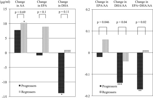
After 8 months of statin therapy, plaque progression was observed in 46 patients (46%), whereas the remaining 55 patients (54%) showed plaque regression. A significant decrease in DHA levels was observed, and EPA levels slightly decreased in the progressors, but this decrease was not observed in the regressors ( Table 2 , Figure 1 ). As a result, serum EPA and DHA levels at follow-up were significantly lower in progressors than in the regressors. Increases in AA levels were observed in the 2 groups. Furthermore, there was a significant decrease in the DHA/AA ratio and EPA+DHA/AA ratio; the EPA/AA ratio slightly decreased in the progressors, while this decrease was not observed in the regressors. Significant differences were observed in changes of the EPA/AA ratio, DHA/AA ratio, and EPA+DHA/AA ratio between the 2 groups ( Figure 1 ).
| Variable | Progressors (n = 46) | Regressors (n = 55) | ||||
|---|---|---|---|---|---|---|
| Baseline | Follow-Up | p Value | Baseline | Follow-Up | p Value | |
| AA (μg/ml) | 126.8 ± 29.4 | 134.7 ± 35.1 | 0.06 | 133.9 ± 28.7 | 144.1 ± 37.0 | 0.02 |
| Mean change (μg/ml) | 7.8 ± 27.6 | 10.2 ± 31.9 | ||||
| DHLA (μg/ml) | 28.1 ± 10.4 | 29.5 ± 11.1 | 0.14 | 31.2 ± 11.4 | 31.6 ± 11.0 | 0.81 |
| Mean change (μg/ml) | 1.5 ± 6.6 | 0.4 ± 11.4 | ||||
| EPA (μg/ml) | 54.2 ± 26.3 | 53.3 ± 22.0 † | 0.79 | 66.2 ± 37.6 | 75.2 ± 43.0 | 0.058 |
| Mean change (μg/ml) | −0.9 ± 24.2 | 8.9 ± 34.3 | ||||
| DHA (μg/ml) | 123.4 ± 47.2 | 109.7 ± 36.6 ∗ | 0.01 | 127.8 ± 51.1 | 128.7 ± 45.7 | 0.9 |
| Mean change (μg/ml) | −13.8 ± 34.7 | 0.9 ± 53.4 | ||||
| EPA/AA | 0.43 ± 0.19 | 0.41 ± 0.19 ∗ | 0.33 | 0.51 ± 0.33 | 0.57 ± 0.38 | 0.07 |
| Mean change | −0.02 ± 0.15 ∗ | 0.05 ± 0.22 | ||||
| DHA/AA | 0.98 ± 0.31 | 0.84 ± 0.28 | <0.0001 | 0.98 ± 0.35 | 0.94 ± 0.35 | 0.28 |
| Mean change | −0.14 ± 0.19 ∗ | −0.04 ± 0.28 | ||||
| EPA+DHA/AA | 1.42 ± 0.46 | 1.25 ± 0.43 ∗ | 0.0009 | 1.49 ± 0.63 | 1.50 ± 0.69 | 0.82 |
| Mean change | −0.17 ± 0.31 ∗ | 0.01 ± 0.43 | ||||

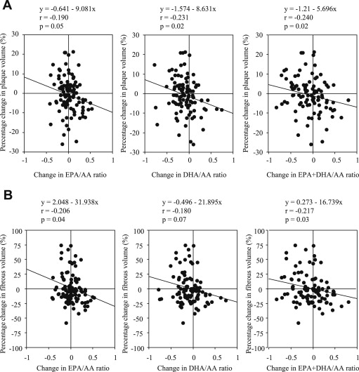
We assessed correlations between percentage change in plaque volume and change in the n-3 to n-6 PUFA ratios. We found that percentage change in plaque volume was negatively and significantly correlated with changes in the EPA/AA ratio, DHA/AA ratio, and EPA+DHA/AA ratio ( Figure 2 ). Furthermore, the percentage change in the fibrous component volume was negatively and significantly correlated with changes in the EPA/AA ratio or EPA+DHA/AA ratio and tended to correlate with changes in the DHA/AA ratio ( Figure 2 ). Percentage changes in fibrofatty, necrotic-core, and dense-calcium components were not correlated with changes in n-3 to n-6 PUFA ratios.

Among n-3 to n-6 PUFA ratios, we used the EPA+DHA/AA ratio in the regression model because this ratio showed the greatest correlation coefficient between n-3 to n-6 PUFA ratios and percentage change in plaque volume or percentage change in fibrous component volume. Univariate and multivariate regression analysis showed that age and change in the EPA+DHA/AA ratio were significantly associated with percentage change in plaque volume or the fibrous component volume ( Table 3 ).


