Echocardiography in Congenital Heart Disease

51 Echocardiography in Congenital Heart Disease


Multipleplane cardiac imaging by echocardiography can noninvasively define the anatomy of the heart and great vessels by delineating the cardiac structures’ configuration, positioning, and spatial interrelations. The information obtained can be used to accurately diagnose and determine prognosis in complex congenital heart disease. With advances in pulsed and color Doppler echocardiography and improvements in the size and capabilities of transducers and other imaging equipment, pediatric echocardiography has gained rapid acceptance. In many pediatric cardiac tertiary care centers, echocardiography is the only diagnostic test performed before neonatal congenital heart surgery. The technology allows real-time three-dimensional (3D) imaging, assessment of myocardial function, and precise definition of cardiac anatomy from the fetal stage through adulthood. For these reasons, echocardiography has become the standard noninvasive diagnostic imaging modality for pediatric cardiology.


Transthoracic multiplane imaging by two-dimensional (2D) echocardiography defines the anatomy of the heart and great vessels. Analysis of each cardiac segment allows complete definition of the configuration, position, and spatial interrelations of cardiac structures. Because tortuous vessels may be difficult to define by a “slice” technology such as echocardiography or MRI, color flow Doppler echocardiography is customarily used to provide a map of blood velocity and direction that complements the 2D image. Small septal defects and fistulous connections may be recognized only by perturbations in blood flow when the anomaly is too small to be visualized clearly. Pulsed and continuous-wave Doppler echocardiography provide excellent time resolution that allows precise quantification of blood velocity. Positional and velocity information can be combined to assess the presence and severity of a valvular obstruction or insufficiency, the position and size of jets associated with septal defects, and abnormal flow in larger vessels in congenital lesions such as anomalous systemic and pulmonary venous return, coarctation of the aorta, and patent ductus arteriosus.


Transesophageal echocardiography (TEE) allows imaging planes different from those obtained in a standard transthoracic study as well as improved visualization of cardiac structures that are difficult to image by transthoracic echocardiography (TTE). Miniaturization of transducer components now allows TEE to be performed in infants as small as 2.5 kg. Posteriorly located cardiac structures are often not well visualized by TTE but are almost always well visualized by TEE. In the older child or a child in whom there are poor transthoracic windows, TEE can also be useful in evaluating congenital heart disease. During cardiac surgery, it may be important to address specific issues by TEE such as anomalous pulmonary venous return, pulmonary vein stenosis, or atrial baffle flows. In addition, an immediate intraoperative or postoperative assessment of the adequacy of surgical repair is possible. While these targeted perioperative examinations do not substitute for a complete preoperative transthoracic evaluation, the availability of TEE has been of great importance in the surgical correction of complex congenital heart disease.


Fetal echocardiography is the newest frontier in pediatric echocardiographic imaging. With the use of transvaginal transducers, detailed fetal cardiac anatomy can be seen as early as 12 weeks of gestation. Transabdominal imaging can be performed by 16 weeks’ gestation, although the optimal time for fetal echocardiography is 18 to 24 weeks. Abnormalities detected then may be important in decisions for further imaging, chromosomal testing, or even pregnancy termination. As with TTE of infants, fetal echocardiography can identify intracardiac anatomy, blood flow across all heart valves, size and orientation of the great vessels, cardiac function, and cardiac rhythm. The order and the windows used in a fetal echocardiogram depend on the fetus’ position, size, and motion, and the amount of fluid in the uterus.


Transthoracic Imaging in Pediatrics


Each pediatric echocardiography laboratory has specific protocols for acquiring a complete study of cardiac anatomy in children. Because patient cooperation is needed, all images may not be obtained using standardized imaging planes in young children and infants. Some centers use conscious sedation for patients under a certain age to ensure uniformity of studies. Another option is “video sedation”: child-friendly videos played to distract the patient and allow time to obtain diagnostic images. As long as clear pictures are obtainable, scanning can be performed with the patient sitting in a parent’s lap, feeding, or even in a stroller. This approach substantially decreases the number of patients who must be sedated.


The protocol for a complete study includes views from the four major echocardiographic windows: parasternal, apical, subxiphoid, and suprasternal. Each window provides the image of the heart from a different angle, allowing multiple, corroborating views of the same structures. The image from each window begins from a standard reference view; then a sweep of the heart is made, first with 2D scanning and then with color Doppler. The color Doppler mapping defines the location for pulsed Doppler scanning in each plane. Once pertinent information is obtained, the transducer is rotated 90 degrees to perform an orthogonal sweep. The sonographer and the interpreting physician can reconstruct multiple 2D images into a 3D representation of cardiac anatomy.


Cardiac function and blood flow are calculated from Doppler mapping and from the 2D images. For example, pulmonary and aortic flow are calculated from the mean velocity and diameter of the vessel at the area of interest as follows:


image


Peak instantaneous gradients are calculated from a simplified Bernoulli equation, using peak flow velocity within the stenotic jet in the following formula, where V is the peak flow velocity measured by spectral Doppler scanning:


image


This gradient is used to estimate pressures in the different cardiac chambers. Several different methods can be used to quantify left ventricular (LV) function. LV fractional shortening (FS) is a measurement of the percentage of change in LV diameter:


image


Ejection fraction is similarly calculated, using measured LV volumes.


Echocardiographic examinations are described below for some common congenital heart lesions, with emphasis on information needed to plan surgical intervention and the best techniques to obtain this information.


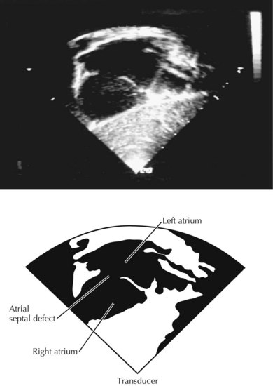
Atrial Septal Defect


TTE is often sufficient to define the size and location of an atrial septal defect (Fig. 51-1). Pulsed and color Doppler echocardiography identify the direction and amount of shunting at the atrial level. Other findings can confirm the presence of a hemodynamically significant shunt. For instance, right ventricular (RV) volume overload can produce diastolic bowing of the ventricular septum to the left during diastole, with the left ventricle assuming an elliptical shape. Partial anomalous pulmonary veins can be identified by 2D echocardiography and color Doppler, and can be corrected at the time of surgery. Echocardiography is usually sufficient to define the anatomy of the atrial septum and pulmonary veins in infants and small children. For older children and adults, TTE may be needed for full anatomic definition. Cardiac catheterization is not usually needed to evaluate atrial septal defects.


Jun 12, 2016 | Posted by in CARDIOLOGY | Comments Off on Echocardiography in Congenital Heart Disease

Full access? Get Clinical Tree

Get Clinical Tree app for offline access