Figure 47.1
Open transthoracic approach. Open transthoracic plication is the traditional approach to treating patients with symptomatic diaphragm eventration or paralysis. Most authors recommend a posterolateral thoracotomy through the sixth to eighth intercostal space (ICS). A variety of techniques for augmentation have been described, including hand-sewn U- stitches, mattress sutures, running sutures, and stapling devices, with or without mesh reinforcement. The direction of the plication (transverse or anteroposterior) is determined by the grossly apparent axis of the eventration. Generally, plication is performed according to a transverse axis (a). Two Babcock forceps raise the slimmed cupola, creating a fold. This fold is fixed at its base with a series of U-shaped nonabsorbable stitches (“flag” plication). The plicated area is folded onto the portion of the diaphragm that appears weaker and fixed close to the intercostal insertion of the diaphragm by one or several rows of sutures, creating a three-layer augmentation at the level of the weakened portion. Mechanical stapling at the base of the fold has been replaced by the U-stitches. Another technique to reinforce the weak portion of the diaphragm is the “accordion” plication, often performed by pediatric surgeons (b). In this technique, mattress sutures are placed in an anteromedial to posteromedial direction, creating the appearance of an accordion. The diaphragm may be plicated with as many rows of sutures as necessary to tighten it. It may be helpful to apply pledgets to the final layers to prevent the sutures from being torn out. If there is a defect or hernia, or the membrane of the diaphragm is very thin, mesh reinforcement may be considered. Significant short- and long-term improvement in dyspnea and respiratory function, as well as patient satisfaction, has been shown after open transthoracic plication. However, thoracotomy and single-lung ventilation are required but may be prohibitive in patients with multiple comorbidities and marginal functional status. a, b, c and a, b refer to the order of surgical steps


Figure 47.2
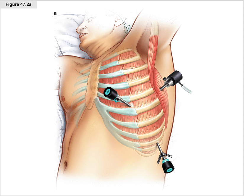
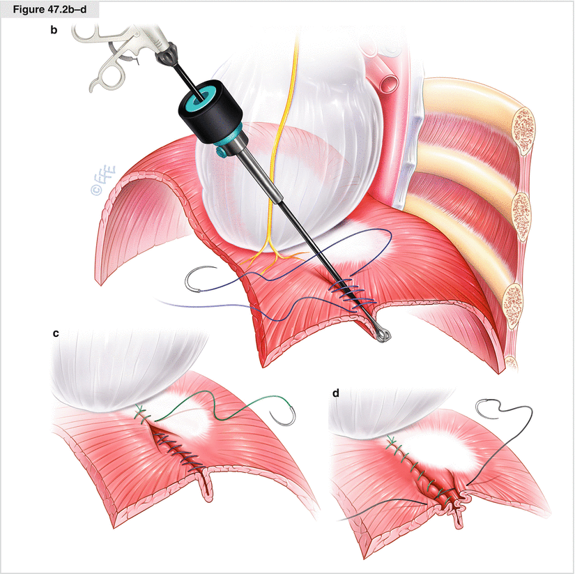
Thoracoscopic approach (VATS). An alternative approach is thoracoscopic plication by VATS, which may be performed with a minithoracotomy using three or four access ports. An example of this surgical technique is shown here. General anesthesia is provided via double-lumen intubation. The patient is placed in the full lateral position indicated for standard posterolateral thoracotomy. Two Thoracoports (10- or 5-mm; Covidien, Mansfield, MA) are placed in the fifth ICS on the posterior axillary line (for the camera) and on the mammary line (for the grasper). A 4- or 5-cm minithoracotomy is made over the ninth ICS on the posterior axillary line for suturing the diaphragm (a). The apex of the eventration is grasped with a Babcock forceps and pushed down toward the abdomen (b). The created transverse fold is sutured with nonabsorbable material beginning at the periphery of the diaphragm close to the minithoracotomy with a running suture back and forth (c). The first continuous suture toward the cardiophrenic angle is placed superficially to avoid injury to the subdiaphragmatic organs. Once at the cardiophrenic angle, the sutures are drawn tight while the forceps used to push down the diaphragm is removed. A row of return stitches is made and tightened with the free end of the first knot. A second back-and-forth series of continuous sutures is placed similarly (d). If there is a defect in the muscular portion or the membrane of the diaphragm is very thin, mesh reinforcement may be considered. With regard to the open approach, several plication techniques have been described (continuous sutures, interrupted sutures, laparoscopic stapling). Thoracoscopic diaphragm plication is an excellent minimally invasive alternative to open transthoracic plication. The disadvantages of VATS are the required single-lung ventilation and the limited workspace

Figure 47.3


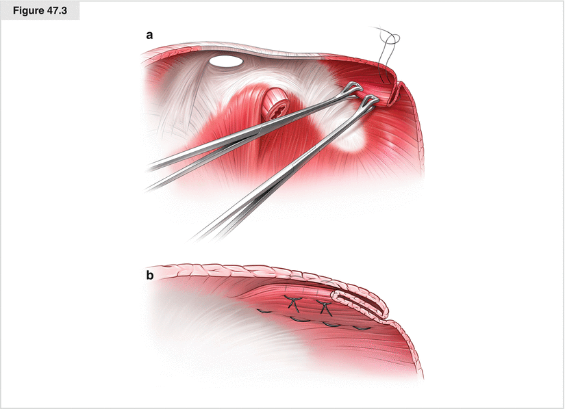
(a, b) Open transabdominal approach. Open transabdominal plication or repair of the diaphragm has been described for unilateral or bilateral diaphragmatic eventration, paralysis, or defects. It is performed over a median or transverse laparotomy. The diaphragm is grasped with two Babcock forceps, and a large transverse fold is drawn downward. The pleat is created with mattress stitches at the base, folded to the anterior circumference, and fixed frontally. A complication may be lung puncture with pneumothorax. Pleuropulmonary adhesions might make this approach unsuitable or even dangerous. Few outcome data are available on the results of open transabdominal plication in adults. Currently, it rarely is used. Advantages may be the laparotomy itself, which generally is less morbid than a thoracotomy; the elimination of the need for single-lung ventilation; and the access it provides to both sides of the diaphragm with one incision. Disadvantages are the difficult access to the posterior portion of the diaphragm and the higher morbidity associated with an open approach
Stay updated, free articles. Join our Telegram channel

Full access? Get Clinical Tree


