Diagnostic Techniques in Vascular Disease

45 Diagnostic Techniques in Vascular Disease


Peripheral vascular disease (PVD) encompasses a broad spectrum of arterial diseases that reduce or alter tissue perfusion. Morphologic changes that narrow the vessel lumen (stenosis) or abnormally enlarge its diameter (aneurysms) cause alterations in flow patterns, thereby leading to ischemic symptoms. Evaluation of PVD always involves advanced imaging. The imaging method of choice depends upon the vessels in question and the end organs and tissue that are affected. Noninvasive imaging techniques include Doppler ultrasound, multidetector CT angiography, and magnetic resonance angiography (MRA). Digital subtraction angiography (DSA) can be utilized to confirm the results of a noninvasive examination and to guide catheter-based therapies. This chapter summarizes the various diagnostic techniques available to evaluate the most commonly encountered occlusive and aneurysmal clinical disorders of the aorta and renal, carotid, and lower and upper extremity arteries.


Etiology and Pathogenesis


A large number of pathologic processes result in arterial occlusive and aneurysm diseases. By far the most common etiology is atherosclerotic plaque formation in the vessel wall leading to luminal narrowing, wall calcification, plaque destabilization and hemorrhage with rupture, thrombosis, and end-organ damage or showering of emboli distally with resulting tissue damage and necrosis. In other circumstances, atherosclerosis leads to weakening of the vessel lumen leading to true aneurysms, defined as a vessel diameter greater than 1.5 times its normal diameter involving all layers. Genetic abnormalities and environmental exposures are almost always contributory in individuals with advanced vascular pathology. Specific genetic abnormalities have been elucidated in Marfan’s syndrome (fibrillin-1), Ehlers-Danlos syndrome, William’s syndrome (hypercalcemia), Loeys-Dietz syndrome (transforming growth factor-β receptor), and homocysteinuria. Thromboangiitis obliterans (Buerger’s disease), which is usually associated with heavy nicotine use, is also seen with PVD. Other risk factors for atherosclerosis (e.g., hypertension, diabetes, lipid abnormalities) all accelerate disease progression and may lead to a need for surgical or percutaneous intervention. Extrinsic mass effects or vessel encasement in the case of popliteal artery entrapment by the gastrocnemius muscle, celiac artery by the diaphragmatic crus (median arcuate ligament syndrome), or renal artery by neurofibromatosis may lead to similar occlusive symptoms. Clinical manifestations depend on the location and severity of the vessel disease and presence of collateral circulation (see Chapter 44 for additional information on the pathogenesis of atherosclerosis).


Lower Extremity Peripheral Vascular Disease


Diagnostic Approach


Examinations used to evaluate the lower extremity arteries are designed to establish the presence and location of disease, quantify severity, and determine temporal progression.


Ankle-Brachial Index and Segmental Pressure Measurements


The ankle-brachial index (ABI) is the simplest baseline examination used for PVD screening. This examination has a high positive predictive value except in diabetics with noncompressible calcified vessels. As an arterial stenosis increases, there is a progressive decrease in the systolic blood pressure (SBP) distal to the stenosis. The decreased blood pressure can be quantified and localized using pneumatic cuffs and either continuous Doppler or plethysmographic sensors. The ABI is determined by measuring SBP in the tibial and brachial arteries. Normally, SBP is amplified in the distal limb by pulse-wave reflection, and the ABI is greater than 1. An ABI of 0.80 to 0.90 is considered to be mildly diminished; 0.50 to 0.80, moderately diminished; and less than 0.50, severely diminished. These values reflect the degree of proximal PVD.


The ABI is simple and inexpensive to use and is an excellent office-based screening tool to identify individuals at risk for limb ischemia who may warrant further investigation if results are abnormal. Pressure measurements at multiple levels along the leg can estimate the location of an arterial occlusion. Blood pressure cuffs are placed on the upper and lower thighs and calves, and a gradient of greater than 10 to 15 mm Hg between adjacent sites suggests a physiologically significant stenosis. Measurements after treadmill exercise may disclose a hemodynamically significant lesion that is unapparent at rest.


Duplex Ultrasonography


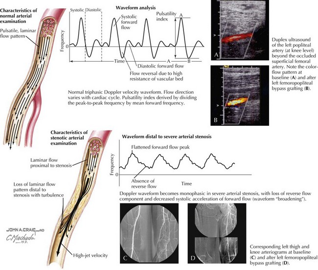
Doppler waveform analysis is an accurate method for defining arterial lesion location and severity (Fig. 45-1). Significant arterial stenoses alter the pattern of flow velocity, as assessed by continuous-wave Doppler analysis. A change in the flow velocity waveform—an increase in peak systolic velocity at the site of the lesion, turbulence, loss of the reverse flow component, or a decrease in pulse velocity distal to the lesion—is diagnostic of a flow-limiting arterial lesion. Color Doppler imaging is able to identify the vessel(s) of interest. A critical stenosis (>50%) is characterized by poststenotic turbulence on color imaging (see Fig. 45-1) and a doubling of the peak systolic velocity on continuous-wave Doppler.


Computed Tomography Angiography and Magnetic Resonance Angiography


Current multidetector or dual-source CT scans with power-injected intravenous contrast provide rapid high-resolution axial scans with imaging of the major vessels throughout the entire cardiovascular system. The temporal spatial and contrast resolution of these scanners to 0.3 mm has allowed improved diagnosis of the location and severity of occlusive and aneurysmal disease, and also can provide information on whether plaques are calcific and whether a thrombus is present. Three-dimensional (3D) curved multiplanar reconstruction, rotational imaging, and postprocessing techniques, such as volume-rendering imaging and stenosis quantification, allow for a thorough analysis of a specific lesion or aneurysm. These outpatient studies have replaced diagnostic angiography in many situations and are completely adequate for targeting interventions, thereby reducing the volume of contrast required and length of examination. CT angiography (CTA) is widely available. The major contraindications to its use are an allergy to iodine (and intravenous contrast) and renal insufficiency. Patients with known or potential dye allergies can be pretreated to reduce allergic reactions, and depending on the degree of renal insufficiency, it is often possible to reduce the risk of worsening renal failure by hydration before and after the procedure. The incidental diagnosis of additional unrecognized disease occurs frequently during the PVD CT scan. Asymptomatic lung, renal, bladder, and other cancers, more common in a population using nicotine, are often discovered during diagnostic CTA. The use of CTA must be weighed against the above complications and the significant radiation exposure dose, especially for younger patients.

Stay updated, free articles. Join our Telegram channel

Jun 12, 2016 | Posted by in CARDIOLOGY | Comments Off on Diagnostic Techniques in Vascular Disease

Full access? Get Clinical Tree

Get Clinical Tree app for offline access