Diagnosis and Treatment of Upper Extremity Vasospastic Disease


Diagnosis and Treatment of Upper Extremity Vasospastic Disease


Santhi K. Ganesh


Vasospasm of the upper extremities is most commonly caused by Raynaud’s syndrome, but it can also result from medications such as ergot compounds, acrocyanosis, livedo reticularis, or intraarterial injections.


Raynaud’s Syndrome


Raynaud’s syndrome is the most common vasospastic disorder seen in clinical practice. Maurice Raynaud in the late 1800s first described this condition as bilateral symmetric gangrene. At the time, it was referred to as Raynaud’s disease. He proposed that vasospasm was caused by sympathetic nervous system overactivity. However, most patients never have manifestations as severe as gangrene, and the entity is now referred to as Raynaud’s syndrome (RS) by most vascular specialists. It is considered primary if there is no underlying disorder and secondary if it is a manifestation of an underlying or systemic condition.


Primary Raynaud’s Syndrome


Primary Raynaud’s syndrome is not life threatening, and affected patients lead relatively normal lives except for symptoms in the hands and fingers and sometimes in the feet and toes. Short-lived vasoconstriction rarely causes irreversible ischemia. Vasospasm can be present for many years without trophic changes of the fingertips, and tissue loss in primary RS is unusual.


The true prevalence of RS is unknown, but in the United States it has been estimated to affect approximately 5% of the population, and the incidence may be increased in populations living in cooler climates. The large majority of patients with RS (89%) have primary RS. The phenomenon affects women more than men in a ratio of 4:1. The typical age of onset is between 10 and 30 years. There is evidence of familial aggregation of RS.


Patients with RS for more than 2 years without evidence of underlying or systemic disease may be classified as having primary RS. Symptoms are often bilateral. β-Blockers aggravate symptoms in 3% to 6% of RS patients owing to their β-adrenergic vasoactive properties.


The typical discoloration of digits has been referred to as the patriotic colors, with white, blue, and red phasic color changes. If vasospasm is severe, there is little or no blood flow to the digits, and they can appear cadaveric white. If vasospasm is moderate, flow may be sluggish, imparting a blue or cyanotic color to the fingers. On rewarming, hyperemia often develops, causing a reddish color in the digits.


Cooling of the hands, parts of the body other than the hands, and the whole body can lead to secondary vasospasm of the fingers in normal persons. These responses may be exaggerated in patients with RS. Vasospasm caused by vasoconstriction in digital arteries and arterioles is a result of increased vascular smooth muscle tone. The increased tone may be caused by α2-adrenergic effects or possibly overactivity of the sympathetic nervous system. In primary RS, there is typically no evidence of arterial occlusive disease. After severe and prolonged disease, mild fibrotic changes have been noted in digital arteries on histopathologic examination.


The major clinical manifestation is the patient’s description of episodes of phasic color changes of the fingers, typically provoked by cold exposure or emotional stress. In addition to discoloration of the fingers, associated symptoms include numbness, paresthesias, and pain or an aching sensation. Typically the fingers are involved, and toes may be affected. Only rarely are toes involved without involvement of the fingers. Approximately 98% of patients report that exposure to a cold environment precipitates symptoms.


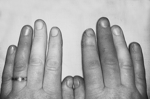
Physical examination is often unrevealing in patients with RS unless they are examined in the midst of an episode, in which case discoloration of the fingers may be present as pallor or cyanosis. The discoloration may be asymmetric (Figure 1). Precipitating an episode by immersing the hand in cold water is inconsistent. An Allen test can show sluggish or mottled return of color to the hand. Livedo reticularis, or the appearance of a mottled or reticular pattern in the skin over the arms and legs, often in a circular pattern, may be seen during an episode of cold exposure in patients with primary RS. In the case of livedo reticularis occurring in primary RS, the findings reverse upon warming of the skin and do not lead to tissue loss or breakdown. Nonreversible livedo reticularis may be observed in other diseases, such as anti-phospholipid syndrome, vasculitis, and atheroembolic or thrombotic disease,


Secondary Raynaud’s Syndrome


Secondary Raynaud’s syndrome may be associated with collagen vascular diseases, hypothyroidism, hyperviscosity syndromes, and many other entities (Box 1). Among rheumatologic diseases, the incidence of RS varies, occurring in approximately 90% of patients with scleroderma and 30% of patients with systemic lupus erythematosus. In secondary RS, particularly with scleroderma and CREST syndrome, manifestations include severe pain, skin ulceration, and tissue infarction, most commonly seen as necrosis of fingertips. In contrast, these findings and tissue loss are rarely observed in primary RS. A positive serologic screen for rheumatologic conditions in an RS patient is highly predictive of the development of a collagen vascular disease. Evidence of arterial obstruction on vascular laboratory testing also predicts digital ulceration in these patients. Secondary RS as a result of other causes, such as hypercoagulable and hyperviscosity states, lead to tissue loss less commonly.


Aug 25, 2016 | Posted by in CARDIOLOGY | Comments Off on Diagnosis and Treatment of Upper Extremity Vasospastic Disease

Full access? Get Clinical Tree

Get Clinical Tree app for offline access