Cutaneous Ulcers in the Neuroischemic Diabetic Foot


Cutaneous Ulcers in the Neuroischemic Diabetic Foot


Ronald Belczyk and George Andros


Ulceration is the most important manifestation of a diabetic foot at risk for amputation. If untreated, the diabetic foot ulcer can enlarge, extend to the deep tissues, and become infected. Deep infection and formation of an abscess can follow and require urgent drainage. Without drainage and intravenous antibiotic therapy, ascending sepsis and septicemia are common sequelae; and if there is associated ischemia, tissue necrosis is virtually inevitable. Because of neuropathy and the resultant loss of protective sensation, this concatenation of complications develops without the patient’s awareness unless a foul odor is detected emanating from the foot. In the late stages, a preemptive major amputation may be unavoidable to save the patient’s life.


Foot ulceration is present in between 3% and 15% of the diabetic population in the United States and most Western countries. Once the ulcer is treated, the looming threat of recurrence is large, in some series greater than 50%. Thus, the best predictor of an ulcer is a previous ulcer. Of all major amputations, 85% are preceded by an ulcer, and for minor amputations there are preexisting ulcers in nearly 100% of cases. Diabetes affects 8.3% of the United States population, nearly 27 million people, and more than 90% have type 2 diabetes. The major complications of diabetes—neuropathy, retinopathy, and nephropathy—occur with approximately equal virulence and ferocity in type 1 and type 2 diabetes. The responsible factor for each complication appears to be sustained hyperglycemia.


Etiology and Pathophysiology


Most ulcers begin because of neuropathy. A small area of the skin is exposed to acute minor trauma such as an abrasion or small laceration. This injury commonly is adjacent to or overlying a bony prominence such as a claw toe, bunion, or Charcot deformity. More often the trauma is repetitive pressure from the simple act of walking. In either instance, an ill-fitting shoe can contribute to constant pressure on the skin. It is this combination of sensory neuropathy that causes the patient to ignore the nascent ulcer, the pedal deformity that often results from associated motor neuropathy, and repetitive trauma that is the core pathophysiology for development and propagation of an ulcer. It is at this early stage that simple interventions like offloading and properly prescribed footwear can allow the ulcer to heal.


Offloading is an essential complement to all forms of therapy. Offloading assists with ulcer healing by decreasing mechanical stress, shear, and trauma. Patient compliance, however, is often problematic because patients in many cases do not have pain and they are not aware that repetitive trauma is occurring. Alternative offloading devices and strategies include non–weight bearing with crutches, walker, wheelchair, or bed rest. However, attempting to keep patients at least partially weight bearing with removable total contact casts, cast walkers, knee walkers, half shoes, and prescription accommodative shoes is the goal.


Care of the skin with lotion to maintain skin softness prevents deep skin cracks, which are often painful over the heels. Dry skin is the product of autonomic neuropathy, which autosympathectomizes the extremities.


Infection may be superimposed on large ulcers or on ulcers as small as a puncture wound, thus complicating management. Likewise, ischemia, depending on its severity, might have almost no effect on ulcer healing or could completely prevent it until pulsatile arterial flow to the foot has been restored. In emerging countries, type 1 diabetes and neuropathic ulcers predominate, whereas in Western industrially advanced countries about 50% of diabetic foot ulcers are neuroischemic in origin so that more complex management strategies, with their increased utilization of medical resources, are routine.


Distribution


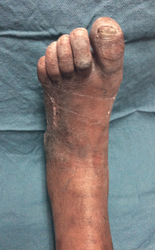
There is a continuum from simple neuropathic to complex neuroischemic ulceration, and both types have similar distributions on the foot. Neuropathy becomes evident in 50% of diabetics who have had the disease for 10 years. The classic claw toe causes the first interphalangeal joint to be arched so that it can rub against the top of the toe box of the shoe and can cause an ulcer. Likewise, the distal aspect of the claw toe points directly downward and strikes the sole of the shoe instead of the pad of the toe, causing the tip of the toe to ulcerate (Figure 1). Ulceration can develop on the plantar surface over any of the metatarsal heads; ulcerations over the second and third are the most common.


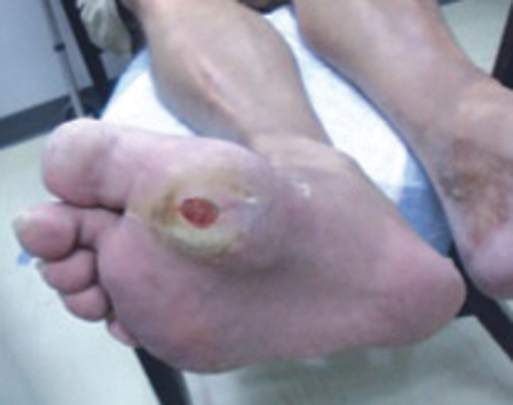
The offloading of the metatarsal high pressure point with appropriate orthotics allows the ulcer to heal, but mal perforans ulcers are prone to recur. Resection of the metatarsal head or the metatarsophalangeal joint offloads the plantar pressure but at the risk of producing transfer lesions under adjacent metatarsal heads (Figure 2). The neuro-osteoarthropathic foot, also known as the Charcot foot, is often characterized by a rocker-bottom appearance. This collapse of the midfoot increases plantar pressure at the point of dislocation. Commonly, the dislocated navicular and medial cuneiform bones produce large ulcers along the plantar medial aspect of the foot and a dislocated cuboid results in a plantar lateral ulceration (Figure 3). Offloading the plantar surface is essential, usually with a total contact cast or its equivalent. When the cast is properly changed on a weekly basis, most Charcot midfoot ulcers can be healed within 3 to 6 months.


Stay updated, free articles. Join our Telegram channel

Aug 25, 2016 | Posted by in CARDIOLOGY | Comments Off on Cutaneous Ulcers in the Neuroischemic Diabetic Foot

Full access? Get Clinical Tree

Get Clinical Tree app for offline access