Conversion to Open Surgical Treatment after Failed Endovascular Aortic Aneurysm Repair


Conversion to Open Surgical Treatment After Failed Endovascular Aortic Aneurysm Repair


Rebecca L. Kelso and Sean P. Lyden


Endovascular aneurysm repair (EVAR) has proved to be safe and effective for the primary treatment of aortic aneurysms. However, endoleaks, endotension, migration, and stent fracture can lead to aneurysm growth and rupture if not treated in a timely fashion. Although transition to newer-generation material and technology has improved the durability of EVAR, complications still occur. Despite the success rate of secondary interventions, patients who are not candidates for an endovascular repair or have unsuccessful interventions require conversion and explant of their endograft. As the length of follow-up increases, studies have also shown that all devices remain susceptible to the risk of removal.


The overall conversion rate is 0% to 9% based on published literature within various EVAR series. The true risk of conversion is difficult to ascertain because many patients are lost to follow-up or report to a different institution for removal. Devices that are placed outside of the instructions for use have been found to carry a higher risk of endograft failure and subsequent removal.


Differences exist between early and late conversions, which are defined as occurring before and after 30 days, respectively. A systematic literature review found that the incidence of early conversion is 0.3% to 5.9%, with a decrease in recent years. For late conversions they found that the incidence averaged 0.4% to 6.3%. In either situation, conversion to open surgery and endograft explant requires careful planning and technical skill.


Indications


The indications for early conversion are often technical from failure of deployment as a result of anatomic or contralateral gate difficulties, inadvertent coverage of renal arteries, or vessel rupture. In these circumstances, conversion is usually related to poor planning, use of devices outside of the instructions for use (IFU), or lack of technical expertise.


The indications for late explant of an aortic endograft are primarily related to device failure. This may be related to one of the many types of endoleaks with or without an enlarging aneurysm sac or migration. With the exception of a type IV endoleak, all types of endoleaks and endotension have been reported causes of aneurysm rupture. The need for explant may also be urgent or emergent as related to graft infection, including aortoduodenal fistulas, or rupture. Limb complications (such as thrombosis caused by stenoses or kinking) can also be a reason for removal when other endoluminal or surgical revascularization options do not exist.


Close monitoring of EVAR patients with secondary interventions has helped identify and, in many cases, successfully treat complications. Not all endoleaks are equal in terms of risk to the patient, and literature supports early management of type I and III endoleaks to prevent aneurysm rupture. Although endovascular salvage of EVAR complications and even recurrent rupture is a viable and often successful option for many patients, conversion is required for those that fail.


Preoperative Considerations


Preoperative evaluation deserves mention for a few factors including overall patient health, as well as preoperative imaging and preparation. The preoperative strategy is not considerably different from other patients undergoing open aneurysm surgery, including those with rupture or infection. Medical optimization of cardiac and pulmonary status is critical because many patients have been deemed high risk before the initial endograft implant.


Knowledge of the type and placement of the graft before explant is also useful and can be found in the patient’s previous medical records as well as determined by plain abdominal film and computed tomography (CT) imaging. Contrast-enhanced spiral CT imaging enables a three-dimensional (3-D) reconstruction, which usually helps identify the cause of endograft failure and thus plan the surgical options, including clamping positions and reconstruction options.


Operative Techniques


At its most basic, explant of an endograft is approached like that of an open aneurysm repair. Technical challenges encountered during removal of endografts include management of suprarenal components, incorporation of stents from endografts into the vessel wall, presence of external stents or barbs, and periaortic inflammation.


In terms of exposure, both retroperitoneal and midline approaches have been used. There are advocates for each approach; however, devices with suprarenal fixation have been more commonly approached by a retroperitoneal approach in most series. Both approaches are equally effective, with the key being the anticipated proximal clamp location. Cases without a suprarenal component and long aortic neck are suitable for infrarenal clamping. Early papers advocated infrarenal clamping, with a temporary release to pull out more proximal portions. However, the ability to temporarily place a higher clamp is of critical importance because releasing the proximal fixation from the aorta can be difficult. In obtaining supravisceral aortic control without an aortoduodenal fistula, it should be the surgeon’s preference and expertise to use a transperitoneal incision with or without medial visceral rotation versus retroperitoneal incision and exposure.


In general, endografts with proximal fixation problems are more likely to be approached from the retroperitoneum with a supravisceral clamp. Once the graft has been removed, the clamp position can be moved a location to limit the renal and visceral ischemic insult. Distal exposure can require the ability to access the external and internal iliac arteries, especially if the entire device is to be removed. An alternative strategy may employ balloon occlusion of the endograft limbs once the aorta is opened and the graft is transected. Some have suggested the use of endoluminal proximal balloon occlusion; however, we find this approach to be cumbersome and unhelpful. To date there is no association between clamp positions (suprarenal vs. infrarenal) and perioperative kidney failure.


Periaortic inflammation can increase the difficulty of the exposure. It has been identified in grafts with both active and passive fixation and grafts with internal and external stents. This finding is unpredictable and not always present—even within similar graft types. Some devices have intimal overgrowth of stents exposed to the vessel wall (e.g., AneuRx, Medtronic, Santa Rosa, CA), and removal of these stents can produce an uncontrolled endarterectomy of the area involved. To aid in removing incorporated portions of endografts we recommend secure control of the aorta above the area in question. The distal device can then be removed in segments when necessary or incorporated into a hybrid open repair when the EVAR device is very adherent to the native vessel wall.


Once the proximal juxtarenal, supravisceral (if needed), and distal control is complete, we recommend opening the aortic sac without clamping unless there is a proximal endoleak. Opening the midportion of the aneurysm sac allows for the evaluation of the positioning of the graft and source of failure. This provides confirmation of the type of endoleak and identifies areas of the graft that are secure in case complete removal is not possible.


Managing and treating the endoleaks without graft explantation may be reasonable when a patient might not physiologically tolerate aortic clamping and complete removal. External suture fixation for type I endoleaks using transaortic to graft sutures has been successfully performed. Some have sutured through felt wrapping for reinforcement of the proximal sealing stent. There are also reports of ligation of lumbar arteries or inferior mesenteric arteries (IMA) responsible for type II endoleaks. Unfortunately, there are also anecdotal reports of continued failures of buttressed grafts with proximal migration as well as development of new types of endoleaks. Choosing this type of open secondary intervention compared to a complete removal with synthetic graft replacement should be carefully weighed in light of the patient’s physiologic status. If the procedure fails, the risk of repeat open surgery would be much higher.


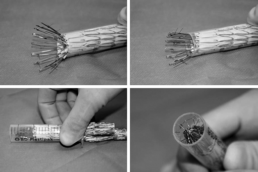
There are many helpful maneuvers to assist in successful removal of the aortic stent graft. The most commonly used is a traditional clamp-and-pull method. In stents with proximal barbs it is helpful to collapse the proximal stent. A technique we have used for removing suprarenal fixation devices with barbs involves collapsing the device into a 20-mL syringe (Figure 1). By cutting off the closed end of a syringe, the syringe cylinder can be then used as a type of short sheath to recapture and separate the proximal stent safely from the aortic wall. For endografts with nitinol stents, iced saline has been suggested to help reduce diameter and ease removal, but we have not found this effective in most circumstances.


Stay updated, free articles. Join our Telegram channel

Aug 25, 2016 | Posted by in CARDIOLOGY | Comments Off on Conversion to Open Surgical Treatment after Failed Endovascular Aortic Aneurysm Repair

Full access? Get Clinical Tree

Get Clinical Tree app for offline access