Congenital Lobar Emphysema
Presentation
A 35-week premature female neonate, weighing 3 kg, is initially discharged home. After 4 days, she is brought to the emergency department by her parents with dyspnea, nasal flaring, tachypnea, and mild cyanosis. She improves slightly with inhalers and humidified oxygen. Breath sounds are decreased on the right side. Endotracheal intubation is not necessary in the initial management.
Differential Diagnosis
The differential diagnosis for severe respiratory distress in a newborn includes congenital heart disease such as vascular rings or slings, mediastinal tumors, bronchogenic cysts, bronchopulmonary dysplasia, congenital lobar emphysema, pulmonary arteriovenous malformations, cystic adenoid malformation, pulmonary sequestration, tracheomalacia, congenital diaphragmatic hernia, and vocal cord malfunction.
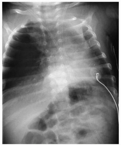
▪ Chest X-ray
Chest X-ray Report
Hyperinflation of the left lung with mediastinal shift to the left side. ▪
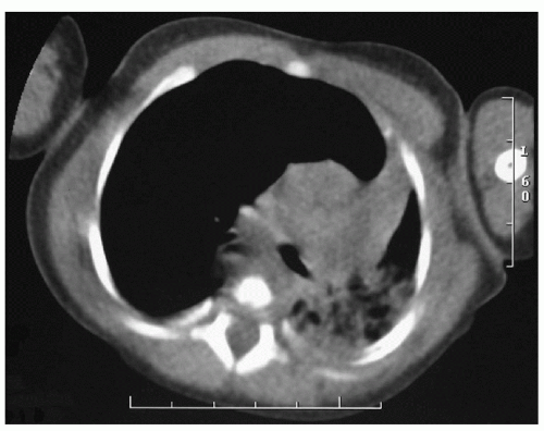
▪ CT Scans


