There have been no optical coherence tomographic (OCT) data directly comparing the pattern of strut coverage between the 2 second-generation drug-eluting stents in the early period. The aim of this prospective study was to evaluate early strut coverage using optical coherence tomography 3 months after Resolute zotarolimus-eluting stent (ZES-R) or everolimus-eluting stent (EES) implantation in de novo coronary artery lesions. A total of 40 patients who were suitable for the OCT procedure and consented to the study protocol were randomized 1:1 to receive either ZES-R or EES. Among these patients, 35 stented lesions (18 ZES-R, 17 EES) in 34 patients were evaluated by optical coherence tomography immediately and 3 months after stent implantation. Neointimal hyperplasia thickness, percentage of uncovered struts, and the proportion of malapposed struts were measured at 1-mm intervals. An uncovered strut was defined as having a neointimal hyperplasia thickness of 0 μm. At the 3-month OCT evaluation, mean neointimal hyperplasia thickness (ZES-R vs EES 74 ± 41 vs 75 ± 35 μm, p = 0.89) and mean percentage of uncovered struts (ZES-R vs EES 6.2 ± 6.9 vs 4.7 ± 5.1%, p = 0.62) were not significantly different between the groups. The percentage of malapposed struts was also similar between the groups (0.7 ± 2.2% for ZES-R and 0.7 ± 1.7% for EES, p = 0.64). Thrombi were documented in 3 stents (1 [5.6%] in a ZES-R vs 2 [11.8%] in EES, p = 0.60). In conclusion, early stent strut coverage on the basis of serial OCT evaluation was comparable between ZES-R and EES 3 months after stent implantation.
Second-generation drug-eluting stents (DES) were developed to address the safety concerns of stent thrombosis while maintaining efficacy similar to that of first-generation DES, and they may offer solutions to the limitations of first-generation DES. The Resolute zotarolimus-eluting stent (ZES-R) (Medtronic Cardiovascular, Santa Rosa, California) and the everolimus-eluting stent (EES) (Xience V; Abbott Vascular Devices, Santa Clara, California) are thin-strut, cobalt chromium–based stents that release zotarolimus from a thin biocompatible coating and everolimus from a thin fluoropolymer-based coating, respectively. In animal studies, endothelial strut coverage at 14 and 28 days after stent implantation was significantly smaller in first-generation DES relative to the more recent EES or ZES. Recent clinical studies have reported more favorable clinical outcomes in patients treated with ZES-R or EES implantation compared with first-generation DESs. We recently reported the results of 9-month follow-up of strut coverage between ZES-R and EES using optical coherence tomographic (OCT) imaging. However, there have been no OCT data directly comparing the pattern of strut coverage between the 2 second-generation DESs in the early period. This type of early OCT data may provide new insight into the status of biologic stability in the early period of new DES struts and information or clues to predict late vascular response patterns. Therefore, this study was designed to compare the early vascular response on the basis of strut coverage 3 months after ZES-R or EES implantation.
Methods
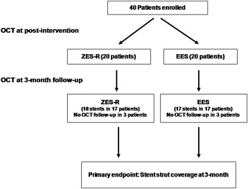
Comparison of Strut Coverage Between Zotarolimus-Eluting Stent and Everolimus-Eluting Stent Using Optical Coherence Tomography 3 Months Following Stent Implantation (COVER OCT II) was a prospective, randomized study to investigate the early vascular healing pattern of second-generation DES using OCT imaging 3 months after stent implantation. From December 2008 to August 2009, a total of 40 patients who were suitable for the OCT procedure and consented to the study protocol were randomized 1:1 to receive either ZES-R or EES, and 35 stents (18 ZES-R and 17 in EES) in 34 patients were evaluated using OCT imaging immediately and 3 months after stent implantation ( Figure 1 ). This trial was performed at 2 university hospitals in Korea. Patients were assigned in a 1:1 ratio using a computer-generated randomization sequence; randomization blocks were created and distributed to the 2 centers.

Inclusion criteria for this study were (1) stable angina or only unstable angina (Braunwald class IB) patients who had de novo lesions with ≥50% diameter stenosis that was related to myocardial ischemia by objective study, or visually estimated stenosis of >70% of the luminal diameter, and (2) native vessel size of 2.5 to 3.5 mm by visual estimation that could be covered by a single stent (multiple lesions in same patient were allowed if lesions existed in different epicardial arteries). Exclusion criteria were (1) unprotected left main coronary artery disease, (2) overlapping stent or bifurcation lesions requiring 2 stents, (3) severely decreased left ventricular systolic function (ejection fraction ≤30%), (4) known intolerance to a study drug, metal alloys, or contrast media, (5) renal insufficiency with baseline creatinine ≥2.0 mg/dl, (6) life expectancy <1 year, and (7) lesions with anatomy unsuitable for OCT imaging using the occlusion technique (proximal vessel size >3.5 mm or proximal lesions <15 mm from the ostium of each artery). The study protocols were separately approved by the institutional review board at each participating institution, and written informed consent was obtained from all patients before participation. The aim of the present trial was to evaluate the early vascular response of 2 second-generation DES as an exploratory study, because there were no data on the expected magnitude of the percentage of uncovered struts 3 months after stent implantation for these DES.
In all patients who signed informed consent, stents were evaluated using OCT imaging immediately after the index procedure and 3 months after stent implantation. Coronary angioplasty was performed according to standard techniques. Adjuvant poststent balloon dilation after OCT examination was performed at the physician’s discretion. Before intervention, all patients were pretreated with a 300- or 600-mg loading dose of clopidogrel within 6 hours of percutaneous coronary intervention. After the intervention, all patients continued to take dual-antiplatelet therapy (aspirin 100 to 200 mg/day and clopidogrel 75 mg/day) during the study period.
A time-domain M2 OCT system (Model M2 Cardiology Imaging System; LightLab Imaging, Inc., Westford, Massachusetts) combined with a 0.014-inch wire-tip imaging catheter (ImageWire; LightLab Imaging, Inc.) was used in this study. The methods of OCT image acquisition were described in a previous report. OCT analysis were performed at an independent core laboratory (Cardiovascular Research Centre, Seoul, Korea). All OCT cross-sectional images were evaluated at 1-mm intervals (every 15 frames) to determine whether stent struts were covered with neointima and apposed to the vessel wall. Among the 701 cross-sectional images, 624 (94.4%) at baseline and 618 (93.4%) at follow-up were included in the analysis. The remaining images were excluded because of poor image quality caused by residual blood, artifacts, or reverberation. A malapposed strut was defined as detachment from the vessel wall that was >117 μm for ZES-R (stent strut thickness 91 μm + abluminal polymer thickness 6 μm + OCT resolution limit 20 μm) or >108 μm for EES (stent strut thickness 81 μm + abluminal polymer thickness 7 μm + OCT resolution limit 20 μm) between the center reflection of the strut and the vessel wall. The thickness of neointimal hyperplasia (NIH) was measured as the distance between the endoluminal surface of the neointima and the luminal surface of the strut, and an uncovered strut was defined as having a NIH thickness of 0 μm. The percentage of uncovered struts was calculated as (number of uncovered struts/total number of struts) × 100. A completely covered stent was defined as a stent with all analyzable struts covered by neointima. A cross section with uncovered struts was defined if ≥1 stent struts was uncovered on cross section, and a cross section with uncovered strut ratio >0.3 was defined when the ratio of uncovered struts to total stent struts per cross section was >0.3. Thrombus was defined as an irregular mass protruding beyond the stent strut into the lumen (>250 μm at the thickest point) with significant attenuation behind the mass. Cross sections with major side branches (diameter ≥2 mm) were excluded from this analysis. The primary end point of this trial was to compare the percentage of strut coverage between ZES-R and EES using OCT imaging 3 months after stent implantation. Quantitative coronary angiographic analysis methods were described in a previous report.
Continuous variables are expressed as mean ± SD and categorical variables as number (percentage). Categorical variables were compared using the chi-square test or Fisher’s exact test. Comparisons of continuous variables were made using paired t tests or Student’s t tests, and the Mann-Whitney U test was applied if the distributions were skewed. A generalized linear mixed model was applied for the patient and cross section analysis, with patient indicator as a random effect and type of stent (ZES-R vs EES) as fixed effect to take into account the clustered nature of the data. Statistical analyses were performed using SAS version 9.1.3 (SAS Institute Inc., Cary, North Carolina). A p value <0.05 was considered to indicate statistical significance.
Results
A total of 40 patients were enrolled in the study and randomized to ZES-R (21 stents in 20 patients) or EES (20 stents in 20 patients) implantation ( Figure 1 ). Baseline clinical and angiographic characteristics between the 2 groups are listed in Table 1 . There were no significant differences between the groups. Among them, 6 patients were excluded because they refused follow-up angiography at 3 months. Adjunct balloon dilation after poststent OCT examination was performed in 10 lesions, 5 (28%) in ZES-R and 5 (29%) in EES (p = 1.00). The rate of malapposed struts significantly decreased from 15.0 ± 5.5% to 1.3 ± 1.4% after adjunct poststent balloon dilation (p <0.001): for ZES-R, from 14.8 ± 6.4% to 0.8 ± 0.8% (p = 0.01), and for EES, from 15.2 ± 5.2% to 1.8 ± 1.8% (p = 0.01).
| Variable | ZES-R (n = 20) | EES (n = 20) | p Value |
|---|---|---|---|
| Age (yrs) | 62.5 ± 9.4 | 66.2 ± 8.3 | 0.19 |
| Men | 14 (70%) | 13 (65%) | 0.74 |
| Acute coronary syndromes | 3 (15%) | 4 (20%) | 1.00 |
| Hypertension | 14 (70%) | 16 (80%) | 0.47 |
| Diabetes mellitus | 9 (45%) | 6 (30%) | 0.33 |
| Dyslipidemia (total cholesterol >200 mg/dl) | 14 (70%) | 12 (60%) | 0.51 |
| Current smokers | 5 (25%) | 2 (10%) | 0.41 |
| Previous myocardial infarction | 2 (10%) | 2 (10%) | 1.00 |
| Number of coronary lesions | 22 | 21 | |
| Target coronary artery | 0.17 | ||
| Left anterior descending | 10 (46%) | 10 (48%) | |
| Left circumflex | 9 (41%) | 4 (19%) | |
| Right | 3 (14%) | 7 (33%) | |
| Type B2 or C lesion | 16 (73%) | 13 (62%) | 0.45 |
| Stent diameter (mm) | 3.1 ± 0.3 | 3.1 ± 0.3 | 0.71 |
| Stent length (mm) | 21.4 ± 5.6 | 19.7 ± 3.2 | 0.24 |
| Maximal pressure (atm) | 14.1 ± 2.0 | 14.1 ± 2.3 | 0.96 |
| Mean reference vessel diameter (mm) | 2.76 ± 0.31 | 2.82 ± 0.30 | 0.52 |
| Lesion length (mm) | 17.9 ± 7.5 | 15.3 ± 3.5 | 0.16 |
| Minimal luminal diameter (mm) | |||
| Preintervention | 1.00 ± 0.39 | 1.21 ± 0.45 | 0.11 |
| Postintervention | 2.54 ± 0.31 | 2.64 ± 0.35 | 0.33 |
| Follow-up (n = 35) | 2.40 ± 0.41 | 2.59 ± 0.33 | 0.14 |
| Acute gain (mm) | 1.54 ± 0.44 | 1.43 ± 0.53 | 0.46 |
| Late loss (mm) (n = 35) | 0.13 ± 0.24 | 0.10 ± 0.15 | 0.66 |
Representative postintervention and 3-month follow-up OCT images of strut coverage for ZES-R and EES are shown in Figure 2 . There were no OCT procedure–related complications. Serial OCT findings are summarized in Table 2 . At the 3-month OCT evaluation, the mean NIH thickness was 74 ± 41 μm in ZES-R and 75 ± 35 μm in EES (p = 0.89), and the percentages of uncovered struts were 6.2 ± 6.9% and 4.7 ± 5.1%, respectively (p = 0.62). The proportion of malapposed struts was also similar between the groups: 0.7 ± 2.2% for ZES-R and 0.7 ± 1.7% for EES (p = 0.64). Thrombi were detected in 3 stents (1 [5.6%] in a ZES-R and 2 [11.8%] in EES, p = 0.60). Of the 35 stented lesions evaluated, 7 lesions (20.0%) were completely covered with neointima: 3 (16.7%) in ZES-R and 4 (23.5%) in EES. The lesions with ratios of uncovered to total stent struts >0.3 were also comparable between the groups ( Table 2 ). There were no deaths, nonfatal myocardial infarctions, target vessel revascularization, or stent thrombosis during the study period.


