Figure 41.1
(a–c) Anatomy of the thoracic duct in the mediastinum. After entering the chest through the aortic hiatus, the thoracic duct ascends on the right side of the vertebral column. In the right lower mediastinum, the thoracic duct is located right in front of the vertebral column, close to the azygos vein and posterior to the esophagus. After its course through the right thoracic cavity, the thoracic duct crosses into the left chest at the T4-T5 level. There it proceeds anterior to the vertebral column behind the aortic arch, where it makes its way to the left jugulosubclavian venous junction. In at least 35 % of cases, the course of the thoracic duct has variations, which must be considered for possible repair in cases of chylothorax. The thoracic duct may be doubled or split into multiple branches, and it often is connected to other vessels via many collaterals. T1–T12 thoracic vertebral
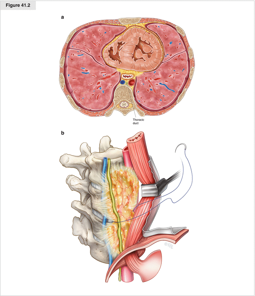
Figure 41.2
(a) Cross-section of the lower mediastinum. The cross-section shows the thoracic duct located in the posterior mediastinum in front of the vertebral body. It is framed by the azygos vein on the right side and the descending aorta on the left. The esophagus appears at the left in front of the thoracic duct and aorta and behind the heart, which fills the anterior mediastinum. (b) Supradiaphragmatic mass ligation via a left-sided approach. In contrast to the right side, where the leakage can be located in most patients, in the left thoracic cavity, sometimes only diffuse lymphatic secretion is seen, and even with the help of preoperatively instilled cream, the site of the leakage cannot be determined. This situation is an indication for supradiaphragmatic mass ligation. After the left inferior pulmonary ligament is released, the lower esophagus is mobilized over a distance of 8–10 cm and retracted to the left side. The mediastinal pleura anterior to the descending thoracic aorta and esophagus is removed to expose the leading edge of the vertebral column. The lymphatic tissue behind the mobilized esophagus between the aorta, the azygos vein, and the leading edge of the vertebral column is dissected, then all remaining fibrofatty tissue superior to the diaphragm between the vertebral bodies, esophagus, aorta, and azygos vein is ligated. Beginning with the ligation at the level of the diaphragm, three or four ligatures of nonabsorbable 3-0 or 4-0 polypropylene sutures are placed over a distance of about 4–6 cm. A complete mass ligation at this site is suitable for stopping chyle leakage from any lymphatic vessel, as well as from unknown accessory ducts. Alternatively, the use of Teflon pledgets to reinforce the sutures used for the mass ligation may be considered
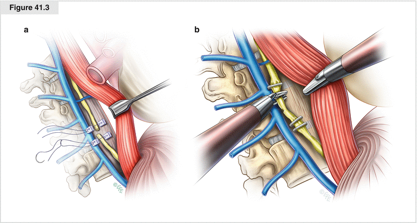
Figure 41.3


(a) Leakage closure by suturing of the thoracic duct. To expose the thoracic duct and gain an overview for finding and repairing a lesion of the duct, the posterior mediastinal pleura between the diaphragmatic hiatus and inferior pulmonary vein is incised. The esophagus is mobilized and retracted off the vertebral bodies anteriorly. After the injured part of the duct is clearly identified and prepared, ligation is performed a few centimeters above and below the leakage site with nonabsorbable sutures, such as 3-0 or 4-0 polypropylene. The stitches are placed close to the thoracic duct above and below the site of injury. Teflon pledgets are placed on both sides of the duct where suturing is to be performed. The sutures are placed through the surrounding tissue close to the duct and through the Teflon pledgets, which also serve as an abutment for a safe knot. To guarantee patency of the duct and to minimize the risk of secondary (suturing-induced) damage, one must avoid placing the stitches directly through the thoracic duct. This suturing procedure is performed on both sides of the lymphatic leak to also stop retrograde leakage. Because sometimes there is more than one lymphatic leakage site and collateral vessels may be present, a final exploration of the upper and lower mediastinum is mandatory to make sure there are no further leaks before the chest is closed. (b) Clipping of the thoracic duct. Clipping of the thoracic duct is an alternative technique that has been applied increasingly because it can be performed safely by videothoracoscopy. After the mediastinal pleura is opened longitudinally, the thoracic duct is exposed, and the lesion is identified, the thoracic duct is clipped on both sides of the leak. Depending on the caliber of the vessel, two to three 5- or 10-mm clips are placed on each side to close the leak safely. To ensure maximum safety, one may also consider applying fibrin glue, especially during a minimally invasive procedure
Stay updated, free articles. Join our Telegram channel
Full access? Get Clinical Tree
