Figure 42.1
Excision and margins. In most cases, general anesthesia is accomplished with a single-lumen endotracheal tube. If associated pulmonary resection is anticipated, single-lung anesthesia is best provided by double-lumen intubation. To minimize postoperative pain and consecutive pulmonary complications, epidural anesthesia is used. After intubation, the patient is placed in a supine or oblique position so that the area of resection is best exposed with all planned options for reconstructive surgery. Single-shot antibiotic prophylaxis should be administered prior to skin incision. To eliminate local recurrence of malignant tumors, the incision is made with at least a 4-cm margin of healthy tissue around the tumor. This approach also should be considered in patients with exulcerated tumors involving the skin or postirradiation soft tissue changes. All previous incisional biopsy sites or surgical scars must be included within the excised en bloc tumor specimen

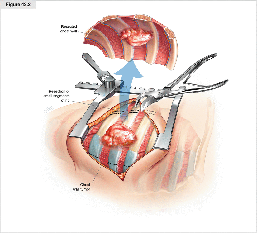
Figure 42.2
En bloc resection of the chest wall. The chest cavity should be entered distal to the tumor as determined by chest CT or MRI. Through thoracotomy or sternotomy, the tumor is palpated within the chest; the lung, hilum, and mediastinum should be palpated for any pathology and to evaluate their resectability. Lymph node involvement and the extent of chest wall involvement must be defined prior to resection. In general, it is not possible to differentiate between chest wall invasion and adhesions reliably by looking at the CT scan. Removal of short rib segments facilitates mobilization of the chest wall segment and provides better visibility of the chest cavity. With resection beginning at the inferior or superior extent, the anterior and posterior margin is taken at each level with the pleura excised. The intercostal muscles are divided along with the pleura, and the posterior portions of the ribs are divided. Hemostasis for the intercostal bundle at each level should be achieved by coagulation or ligation

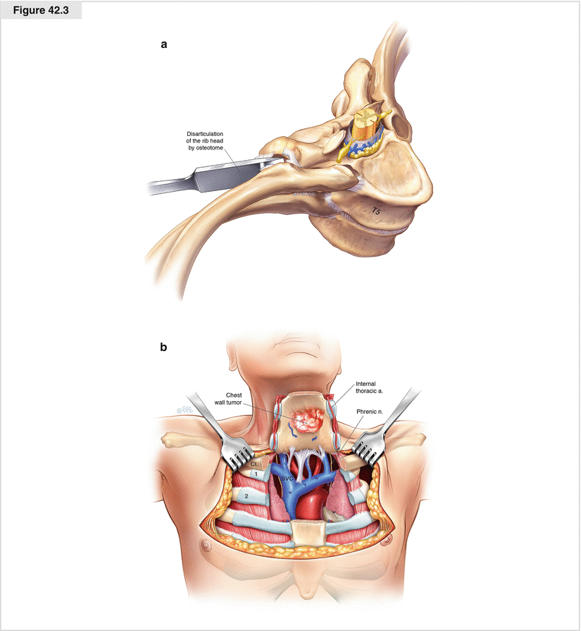
Figure 42.3


Disarticulation from the posterior rib to the transverse process. The patient must be placed at a 45° anterior oblique angle in a lateral position with an axillary roll under the downside to allow access to the dorsal costispinal junction. If disarticulation of the ribs from the transverse process is required to obtain negative margins, the paraspinous ligament must be mobilized from the spine and the cartilaginous junction between the neck of the ribs and transverse processes needs to be incised with either electrocautery or an osteotome. As the pleura from within the chest is incised, the osteotome is directed anteriorly toward the vertebral body and thus away from the spinal canal by lifting the head of the rib. The ribs should be disarticulated gently to avoid tearing the dural sheath and to prevent the resultant cerebrospinal fluid leakage. Intercostal bundles must be ligated at the level of the neural foramen. This needs to be done with care; a transfixion ligature may help prevent slippage with bleeding or opening of the epidural space. Only bipolar electrocautery should be used near the spinal foramen. Bleeding from epidural veins is controlled with bipolar electrocautery, and temporary compression with application of fibrin foam frequently is adequate. An accidental dural lesion should be sutured and covered by placing a small flap of paraspinal muscle. Once the chest wall bloc is mobilized, the attached underlying invaded lung parenchyma must be resected. In most cases involving the primary chest wall, this may be done via wedge resection or segmentectomy. In patients with secondary chest wall tumors by invasion of primary lung cancer, lobectomy with concomitant systemic mediastinal lymph node dissection is advised. (a) Disarticulation from the posterior rib to the transverse process. T5 5th thoracic vertebra. (b) Sternal resection for a chest wall tumor. SVC superior vena cava, CL clavicula, C11st, C2 2nd. rib
Stay updated, free articles. Join our Telegram channel

Full access? Get Clinical Tree


