Cerebrovascular Disease


Cerebrovascular Disease


Susan L. Hickenbottom

Gilbert R. Upchurch Jr.

James C. Stanley

Lewis B. Morgenstern


EPIDEMIOLOGY AND USUAL CAUSES OF CEREBROVASCULAR DISEASE

Stroke is a clinical syndrome characterized by rapidly developing signs or symptoms, or both, of focal neurologic dysfunction with no obvious cause other than vascular origin. Therefore stroke includes both ischemic and hemorrhagic cerebrovascular events. Transient ischemic attack (TIA) is an abrupt, focal loss of neurologic function caused by temporary ischemia, classically defined as lasting less than 24 hours. However, modern imaging techniques have demonstrated that deficits lasting more than a few hours usually result in irreversible cerebral infarction and that most TIAs typically last less than 15 minutes (1). For the same reason, the term reversible ischemic neurologic deficit (RIND), referring to symptoms lasting more than 24 hours but less than 7 days, has been abandoned. The colloquial term cerebrovascular accident (CVA) is also considered outdated, as it implies a fortuitous origin, whereas stroke is usually caused by a well-defined etiopathogenic mechanism.

In 1998, the American Heart Association (AHA) estimated that more than 600,000 strokes and 160,000 deaths of stroke occurred in the United States, which makes stroke the third leading killer of Americans and the leading cause of long-term disability among adults (2). More recently, a multiracial study performed in the Cincinnati metropolitan area found that more than 730,000 first-ever and recurrent strokes occur each year in this country (3). Indeed, stroke risk is much greater in African Americans (4) and Mexican Americans (5), suggesting the need for particular prevention efforts in these populations. Approximately 85% of strokes are ischemic, 10% are caused by parenchymal intracerebral hemorrhage (ICH), and 5% are secondary to subarachnoid hemorrhage (SAH).

Caring for patients with stroke is an expensive undertaking. Recently, projections for the cost of stroke in the first half of the 21st century exceeded 1.5 trillion dollars (6). Loss of earnings is the single biggest contributor, but rehabilitation and nursing home costs add considerably to the total. The cost of informal care giving is often underappreciated (7).

Stroke is not a single disease process but a heterogeneous group of disorders with similar clinical presentation, and thus stroke can have many different underlying causes. The usual causes of ischemic stroke are outlined in Table 31.1. As with ischemic cardiovascular disease, the most common cause of ischemic cerebrovascular disease is atherosclerosis. Risk factors for ischemic stroke include increasing age, male gender, family history of cardiovascular or cerebrovascular disease, hypertension, diabetes, hyperlipidemia, cigarette smoking, atrial fibrillation, carotid artery stenosis, and history of stroke, TIA, or myocardial infarction (8). Other potential risk factors include excessive alcohol intake, sedentary lifestyle, hyperhomocystinemia, hereditary or acquired hypercoagulable states, and obstructive sleep apnea. The usual causes of hemorrhagic stroke (both ICH and SAH) are presented in Table 31.2.


TABLE 31.1. Usual causes of ischemic stroke


Large-artery atherothromboembolism (˜50%)


Thrombosis in situ


Artery-to-artery embolism


Small-vessel atherothromboembolism (˜15%)


“Lacunar” disease associated with hypertension and diabetes


Cardioembolism (˜25%)


High risk


Nonvalvular atrial fibrillation


Rheumatic heart disease


Prosthetic cardiac valves


Infective endocarditis


Moderate risk


After myocardial infarction


Aortic arch atheroma


Dilated cardiomyopathy


Patent foramen ovale/atrial septal aneurysm


Other, less common causes (˜10%)


Arterial dissection


Arteritis, including primary angiitis of the central nervous system and arteritis associated with underlying infectious or immune diseases


Arteriopathies, including moya-moya disease


Hereditary or acquired hypercoagulable states, including polycythemia vera, disseminated intravascular coagulation, thrombotic thrombocytopenic purpura, dysfibrinogenemia, antiphospholipid antibody syndrome, sickle cell disease, factor V Leiden mutation, prothrombin 20210 mutation, protein C/protein S deficiency, antithrombin III deficiency


Of course, the most likely cause of stroke in a given patient depends on the patient’s age, race, ethnicity, and the spectrum of risk factors. In addition, more than one cause of stroke may be present in the same patient.


PRESENTING SIGNS AND SYMPTOMS

The presenting signs and symptoms of stroke are entirely dependent on the location of brain tissue involved in the vascular process.
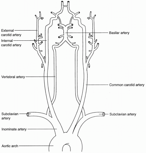
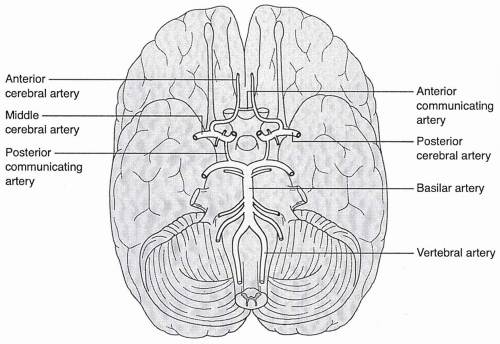
 Figures 31.1 and 31.2 provide a review of the extracranial and intracranial vascular anatomy involved in cerebrovascular circulation. 
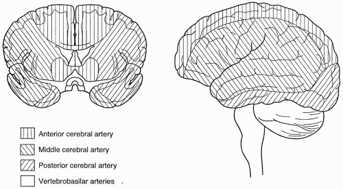
Figure 31.3 outlines the vascular territories of the cerebral circulation. Stroke syndromes have classically been divided into anterior and posterior circulation distributions, on the basis of presenting signs and symptoms, and further subdivided according to the presumed arterial involvement referable to the signs and symptoms. These arterial syndromes and their typical signs and symptoms are outlined in Table 31.3.


TABLE 31.2. Usual causes of hemorrhagic stroke


Intracerebral hemorrhage (10% of all stroke)


Hypertension (>60%)


Bleeding diathesis (5%-20%)


Vascular malformation (4%-8%)


Aneurysm (3%-4%)


Neoplasm (2%-7%)


Other causes: cerebral amyloid angiopathy, drug-related, cerebral venous occlusive disease, hemorrhagic transformation of ischemic arterial infarction, arteritis/arteriopathies, trauma, after carotid endarterectomy or other neurosurgical procedures, postmyelography


Subarachnoid hemorrhage (5% of all stroke)


Trauma


Aneurysm (80%-90% of nontraumatic subarachnoid hemorrhage)


Other causes: vascular malformation, arterial dissection, drug related, cerebral venous occlusive disease, bleeding diathesis, arteritis/arteriopathies


Although stroke signs and symptoms are helpful in localizing the stroke to a specific region of the brain or arterial territory, it is important to remember that the clinical presentation typically does not provide any information about stroke etiology. Headache, vomiting, and decreased level of consciousness are somewhat more common with hemorrhagic stroke, but ischemic stroke can occur with similar symptoms, especially if the vertebrobasilar arterial system is involved or if a hemispheric stroke is large enough to cause mass effect and increased intracranial pressure. Likewise, thrombotic and embolic strokes cannot be distinguished from each other merely on the basis of presenting signs and symptoms.


FIGURE 31.1. Pictorial representation of the arterial supply to the brain.


DIFFERENTIAL DIAGNOSIS AND HELPFUL TESTS FOR THE DIAGNOSIS OF STROKE


Differential Diagnosis of Stroke

The differential diagnosis of stroke includes various other neurologic and medical entities. Keys to distinguishing stroke from other diagnoses include its acute onset (as opposed to subacute onset or chronic progression) and the focality of the presenting neurologic signs and symptoms. In most cases, the correct diagnosis can be established by careful history documentation, physical examination, and diagnostic testing. More recently, however, it has become especially important to be able to diagnose acute ischemic stroke (AIS) rapidly and accurately since the U.S. Food and Drug Administration (FDA) approved the use of tissue plasminogen activator (alteplase) for thrombolysis in acute ischemic stroke in 1996 (see Treatment section). Table 31.4 lists the differential diagnoses for acute ischemic stroke and diagnostic tests that can be used to help confirm this diagnosis.


Emergency Diagnostic Evaluation of Acute Stroke

The majority of patients with acute stroke should be initially evaluated in the emergency department. After attention to the
issues of oxygenation and hemodynamic stability, a medical history and physical examination should focus on specific stroke risk factors and causes, followed by clinical localization of the ischemic territory (Table 31.3). Rapid determination of the blood glucose level should be made to rule out hypoglycemia or hyperglycemia as the cause of the neurologic deficit. Laboratory studies, including a complete blood cell count and
measurement of electrolytes, glucose, and coagulation parameters, should be obtained. A toxicology screen should be ordered for young patients and any others suspected of illicit drug use. Electrocardiography is needed to assess for evidence of arrhythmia or cardiac ischemia. Emergency computed tomography (CT) is necessary to identify ICH, SAH, or early signs of cerebral ischemia. CT appearances of AIS, ICH, and SAH are demonstrated in Fig. 31.4. It is important to remember that initial CT scanning in AIS may be normal or may reveal only subtle, early signs of cerebral ischemia. A sample emergency department protocol for the initial evaluation and management of acute stroke is provided in Table 31.5.


FIGURE 31.2. Inferior view of the brain: arterial supply to the brain with attention to the circle of Willis.


FIGURE 31.3. Coronal and lateral views of the vascular territories of the brain.


TABLE 31.3. Presenting signs and symptoms of stroke by vascular distribution


ARTERY


CLINICAL FEATURES


Anterior circulation


ICA/MCA


Ipsilateral monocular vision loss (amaurosis fugax) ±Contralateral weakness or sensory changes Contralateral weakness, sensory changes (more severe in face and arm than in leg) ±Contralateral visual field deficit Aphasia (dominant hemisphere) Neglect and other visuospatial difficulties (nondominant hemisphere)


ACA


Contralateral weakness, sensory changes (more severe in leg than in face and arm) Personality changes (disinhibition, lack of motivation, disinterest)


Posterior circulation


PCA


Contralateral visual field deficit ± other visual phenomena


VB


Ipsilateral cranial nerve deficits ± ataxia Contralateral or bilateral weakness or sensory changes Contralateral or bilateral sensory changes Diplopia Dysarthria Dysequilibrium or vertigo (not in isolation) Altered level of consciousness


ACA, anterior cerebral artery; ICA, internal carotid artery; MCA, middle cerebral artery; PCA, posterior cerebral artery;


VB, vertebrobasilar system.


Emergency imaging studies may be useful for some treatment decisions. Magnetic resonance imaging (MRI), including diffusion-and perfusion-weighted imaging, may be used to detect early ischemia and to
identify salvageable penumbral tissue that could be targeted with thrombolytic or neuroprotective therapies. Similarly, CT perfusion imaging can also estimate the territory at risk and help guide interventions. CT angiography (CTA) and MR angiography (MRA) are excellent tools for lesion localization. As mentioned later, if a patient is eligible to receive intravenous rt-PA, the only imaging study indicated is a noncontrast head CT to exclude blood. The other imaging tools add time and may delay intravenous rt-PA administration.


TABLE 31.4. Differential diagnosis of acute ischemic stroke


DISORDERS THAT MIMIC ACUTE ISCHEMIC STROKE


DIAGNOSTIC TOOLS


Intracerebral hemorrhage


CT/MRI


Subarachnoid hemorrhage angiography


CT, lumbar puncture, cerebral


Subdural/epidural hematoma


History of trauma, CT/MRI


Structural lesion (e.g., neoplasm)


CT/MRI


Hypoglycemia or hyperglycemia


Fingerstick glucose measurement


Other metabolic derangements


Routine chemistry studies


Seizure


Clinical history, EEG


Complicated migraine


Clinical history


Conversion disorder


Clinical history, psychiatric evaluation


CT, computed tomography; EEG, electroencephalography; MRI, magnetic resonance imaging.


FIGURE 31.4. Computed tomographic appearance of acute ischemic stroke (A), intracerebral hemorrhage (B), and subarachnoid hemorrhage (C). Acute ischemic stroke may demonstrate no or only minimal changes on computed tomograms in the acute setting.


FIGURE 31.4. (continued)


TABLE 31.5. Emergency department protocol for the initial management of presumed acute ischemic stroke


1.


Obtain vital sign measurements, including temperature, pulse, blood pressure, and oxygen saturation; continue to monitor every 15 min


2.


Begin continuous cardiac and oxygen-saturation monitoring


3.


Ensure adequate airway/respiratory status


a.Intubate and initiate mechanical ventilation if necessary


b.Otherwise, begin oxygen at 2 L/min via nasal cannula


4.


IV access: 0.9 normal saline at 100 mL/hr if no CHF; saline lock in opposite arm


5.


Stat laboratory studies


a.Serum glucose (may be measured at bedside)


b.Complete blood cell count with platelet count


c.Chemistry profile


d.Coagulation studies (prothrombin time, activated partial thromboplastin time)


e.Urine pregnancy test for women of childbearing age


i. Urine toxicology screen


6.


Establish patient’s weight (measure or estimate)


7.


Obtain i.v. pump for possible infusion therapy


8.


Order stat head CT without contrast material


9.


No aspirin or other antiplatelet agents, heparin, or warfarin to be given to potential thrombolytic therapy recipients


CHF, congestive heart failure; CT, computed tomography; i.v., intravenous.

Only gold members can continue reading. Log In or Register to continue

Stay updated, free articles. Join our Telegram channel

Aug 18, 2016 | Posted by in CARDIOLOGY | Comments Off on Cerebrovascular Disease

Full access? Get Clinical Tree

Get Clinical Tree app for offline access