Celiac, Hepatic, and Splenic Artery Aneurysms


Celiac, Hepatic, and Splenic Artery Aneurysms


John E. Rectenwald and James C. Stanley


Although uncommon, splanchnic artery aneurysms are being recognized with increasing frequency with the widespread use of advanced imaging techniques. True aneurysms and pseudoaneurysms of the visceral branches of the aorta remain a potential source of significant vascular morbidity and mortality. Management of these aneurysms is evolving to include a more important role for endovascular therapy. Aneurysms involving the celiac, hepatic, and splenic arteries account for more than 80% of all reported splanchnic artery aneurysms.


Celiac Artery Aneurysms


Clinical Presentation


Celiac artery aneurysms account for 4% of all splanchnic artery aneurysms. The mean age at the time of diagnosis is 56 years. Men and women appear to be equally affected. Aortic aneurysms affect nearly 20% of these patients, and approximately 40% have other splanchnic aneurysms. Half the celiac artery aneurysms reported before 1950 were mycotic, although infectious lesions have been uncommonly encountered in recent years.


Most celiac artery aneurysms appear secondary to medial degeneration. Arteriosclerosis is a frequent finding, but, as in the case of other splanchnic artery aneurysms, it is considered a secondary process. Celiac artery aneurysms are usually saccular and involve the distal trunk of the artery. Some evolve from poststenotic dilatations caused by intrinsic occlusive disease or median arcuate ligament compression of the proximal celiac artery.


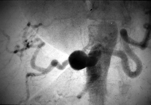
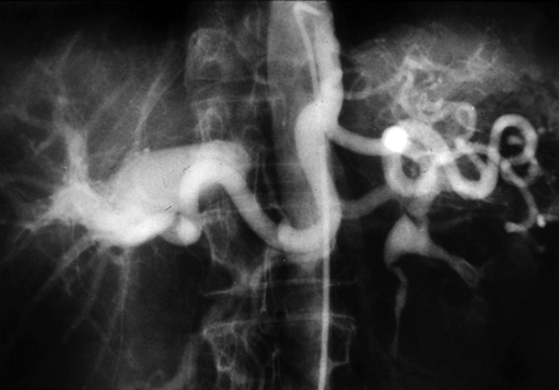
Currently, most celiac artery aneurysms are asymptomatic or are associated with vague abdominal discomfort. They are often first recognized as incidental findings on computed tomography or other imaging for nonvascular diseases (Figure 1). Some patients, however, have symptoms, with abdominal or back pain being the most common manifestation in these cases. Less common presentations are early satiety, jaundice, and gastrointestinal bleeding. Rupture reportedly occurs in 13% of these celiac artery aneurysms, is usually intraperitoneal, and carries a mortality of 50%. Intervention is warranted for all symptomatic celiac artery aneurysms and for bland aneurysms exceeding 2 cm in diameter.


Management


Open surgical procedures are the most common intervention for celiac artery aneurysms unless a prohibitive operative risk exists. Most nonruptured aneurysms can be treated through an abdominal approach, although in the presence of acute expansion or rupture, a thoracoabdominal incision may be favored. Arterial reconstruction of the celiac trunk is preferred following aneurysmectomy, although simple aneurysmal exclusion with ligation of entering and exiting branches may be performed in selected patients. If the latter is undertaken, the foregut collateral blood flow to the liver must be sufficient to prevent severe hepatic ischemia. When such is not the case, an aortoceliac or aortohepatic artery bypass should be undertaken. Successful outcomes of open surgical therapy are greater than 90%.


Endovascular treatment of celiac artery aneurysms is not often performed because of the need to occlude the hepatic, splenic, and left gastric arteries, as well as the inferior phrenic arteries in some cases. Nevertheless, successful endovascular therapy of these aneurysms has been reported and has a role, especially in patients at high risk for open surgery.


Hepatic Artery Aneurysms


Clinical Presentation


Hepatic artery aneurysms account for 20% of all reported splanchnic artery aneurysms. Hepatic artery aneurysms are usually solitary; they are extrahepatic in nearly 80% of cases and intrahepatic in 20%. Arteriosclerosis is considered a secondary event.


They are relatively common in current practice. Traumatic etiologies account for almost half of the reported hepatic artery aneurysms involving the intrahepatic arterial branches. This likely relates to the increased incidence and detection of pseudoaneurysms of the intrahepatic arterial branches resulting from percutaneous diagnostic and therapeutic biliary tract procedures and the computed tomography (CT) recognition of small parenchymal pseudoaneurysms following blunt abdominal trauma.


Most extrahepatic aneurysms are true aneurysms with pathologic changes suggesting medial degeneration. Atherosclerotic changes appear to be secondary. Men accounted for 55% of patients with reported hepatic artery aneurysms since the turn of the century, and the mean age at presentation is 52 years. More than a third of patients with these aneurysms have other splanchnic artery aneurysms. Nontraumatic and nonmycotic aneurysms are most often discovered during the sixth decade of life.


Less common causes of hepatic artery aneurysms include mycotic aneurysms, vasculitides, and inflammatory aneurysms associated with acute pancreatitis or cholecystitis. Connective tissue arteriopathies, such as periarteritis nodosa, have also been incriminated as a cause of occasional macroaneurysms involving the hepatic vessels. Mycotic aneurysms associated with sepsis, often related to intravenous drug abuse and endocarditis, are relatively uncommon.


Most hepatic artery aneurysms are asymptomatic and are discovered incidentally during imaging for other illnesses (Figure 2). Those aneurysms that are symptomatic produce right upper quadrant and epigastric pain in half the patients. Acute expansion of hepatic artery aneurysms can cause severe abdominal discomfort, similar to that of acute pancreatitis. Large aneurysms have been reported to be a rare cause of obstructive jaundice.


The incidence of extrahepatic hepatic artery aneurysm rupture remains ill defined, but the current rupture rate approaches 20%. Rupture of iatrogenic intrahepatic aneurysms has occurred in more than half of the cases from institutions reporting large numbers of this subgroup of aneurysms. Bleeding from ruptured hepatic artery aneurysms occurs equally into the biliary tract and into the peritoneal cavity. In the case of the former, hemobilia is often evident, manifest by biliary colic, hematemesis, and jaundice. Chronic gastrointestinal hemorrhage is an uncommon but recognized sequela of aneurysm rupture into the biliary tract. Intraperitoneal bleeding is usually associated with false aneurysms caused by periarterial inflammatory processes eroding into the hepatic vessels. Multiple nonarteriosclerotic aneurysms have an apparent greater risk of eventual rupture. The reported overall mortality rate attending aneurysm rupture approaches 35%, although recent experience suggests that rupture-related mortality is half that figure.


Management


Operative intervention for hepatic artery aneurysms should be considered in all but very high risk patients. Treatment should be individualized depending on size and location of the aneurysm. Endovascular or open surgical treatment is recommended for all symptomatic aneurysms and all asymptomatic saccular hepatic aneurysms exceedingly 2 cm in diameter. Expanding intrahepatic aneurysms and pseudoaneurysms of the extrahepatic arteries warrant operative intervention.


Percutaneous transcatheter obliteration of hepatic artery aneurysms with balloons, coils, or various types of particulate matter, including thrombin, is preferred over an open surgical intervention. However, transcatheter embolization may be only transiently successful, and repeated embolization or eventual open surgical therapy may be required to adequately treat certain patients. Endovascular stent graft exclusion of select aneurysms avoids some of the limitations accompanying embolization alone but often results in occlusion of the stented artery.


Common hepatic artery aneurysms in the past were usually treated by an open aneurysmectomy, or aneurysm exclusion, without arterial reconstruction. Exposure is generally accomplished through a generous bilateral subcostal incision, occasionally employing a superior midline extension to improve exposure. Vascular control is preferentially obtained by isolating and clamping the inflow and outflow vessel of the aneurysm. However, large aneurysms or significant associated local inflammation can make such dissection hazardous. In these cases, it may be safest to obtain vascular control from within the aneurysm with balloon catheters while briefly clamping the supraceliac aorta to minimize blood loss until the occluding catheters can be placed.


If hepatic blood flow appears compromised after hepatic artery occlusion, a direct vascular reconstruction should be undertaken with a prosthetic or autologous graft. Hepatic ischemia is most likely to accompany treatment of aneurysms involving the proper hepatic artery or its extrahepatic branches and, whenever possible, arterial reconstruction of these vessels is favored following aneurysmectomy. Casual ligation of extrahepatic branches to control bleeding from intrahepatic aneurysms can cause liver necrosis, and in this setting hepatic territory resection may be necessary.

Stay updated, free articles. Join our Telegram channel

Aug 25, 2016 | Posted by in CARDIOLOGY | Comments Off on Celiac, Hepatic, and Splenic Artery Aneurysms

Full access? Get Clinical Tree

Get Clinical Tree app for offline access