Celiac Artery Compression Syndrome


Celiac Artery Compression Syndrome


Guillermo A. Escobar and James C. Stanley


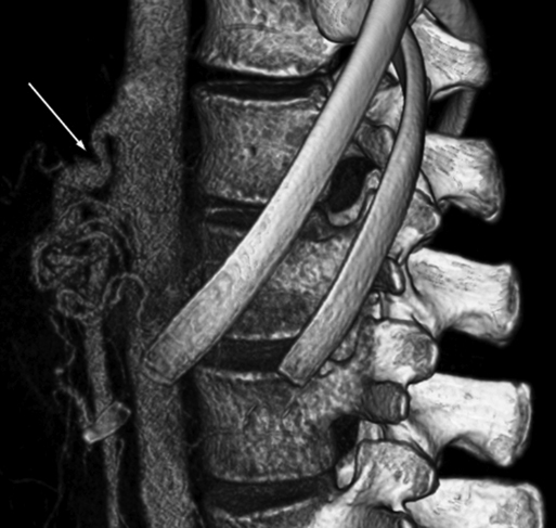
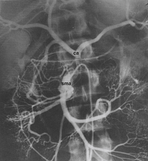
Celiac artery compression syndrome was first described in the 1960s. It is a consequence of extrinsic compression by the median arcuate ligament of the celiac artery and surrounding neural tissues (Figure 1). Several synonyms have been applied to this condition, including celiac axis syndrome, celiac band syndrome, Dunbar’s syndrome, and the median arcuate ligament syndrome. Controversy surrounds the existence of this syndrome because many asymptomatic persons have evidence of celiac artery compression, and caution is appropriate in assigning clinical relevance to the mere anatomic presence of a celiac artery narrowing.


Clinical Manifestations


The classic presentation of the celiac artery compression syndrome is that of a young, thin female patient who has postprandial abdominal pain, food fear, and weight loss suggesting chronic mesenteric ischemia. Most clinical series report a 2:1 predominance of women in their third decade of life, although this disorder also has been described in patients as young as 3 years of age.


Abdominal discomfort is most often periumbilical and epigastric, occurring shortly after eating and lasting 2 to 4 hours. Nausea can accompany the pain, but emesis is uncommon. Although positional precipitation of the discomfort has been reported, it is not a consistent manifestation of the syndrome. An upper abdominal bruit that varies with respiration that is more pronounced with fixed expiration is a common finding. Abnormalities in motility or absorption have not been documented in these patients.


Anatomic Basis of Arterial Entrapment


Median arcuate ligament entrapment of the celiac artery as an anatomic finding dates to 1917, long before the clinical peculiarities attending it were described in the 1960s. Minor indentations along the superior border of the celiac artery by the ligament affect approximately 40% of the adult population. In 15% of cases, more severe narrowings can result in celiac artery occlusion and the development of extensive gastroduodenal and pancreaticoduodenal collateral vessels between the superior mesenteric artery (SMA) and celiac artery circulations (Figure 2).


Three factors appear to contribute to the celiac artery narrowings. First is an abnormally low-lying fibrous median arcuate ligament, which compresses and kinks a normally located celiac artery (Figure 3). Second, the celiac artery can originate higher than usual in the presence of a normally positioned median arcuate ligament. In regard to these two scenarios, both female patients and thin patients have been found to have atypically low-lying diaphragms as well as high origins of their celiac artery. Third, an excessive collection of constricting neural and fibrous tissue can entrap and narrow the celiac artery origin.


An early report from the University of Michigan revealed that the mean level at which the median arcuate ligament crosses the aorta is at the junction of the middle and lower third of the first lumbar vertebra. Given that the celiac artery’s origin is between the lower third of the 12th thoracic vertebra and middle third of the first lumbar vertebra, the potential exists for its compression at the aortic hiatus. The inconsistent location of the celiac artery’s origin is the result of its variable caudal descent during embryonic development. In support of its more cephalad location is the observation that the inferior phrenic arteries arise from the celiac artery, rather than from higher on the aorta, in 52% of the normal population. The mean origin of the SMA is also at the lower third of the first lumbar vertebra, and its close proximity to the celiac artery and aortic hiatus explains its occasional narrowing in patients with the clinical syndrome.


Intraoperative findings invariably reveal a distinctly fibrous median arcuate ligament compressing the celiac artery. Some have suggested that the rigid character of this tissue may be prerequisite to developing clinical symptoms. Ganglionic compression of the celiac artery is also common, and an intermingling of celiac plexus fibers and elements of the aortic hiatus is often evident in biopsies of the median arcuate ligament. In some patients, abnormal blood flow within the celiac artery leads to secondary intimal fibroplasia and a further narrowing of the already compressed vessel.


Physiologic Consequences of Arterial Entrapment


The hypothesis that the primary derangement in the median arcuate ligament syndrome involves purely intestinal ischemia has little objective support. The celiac artery, SMA, and inferior mesenteric artery all contribute to the splanchnic circulation, and the large network of collateral channels between these vessels are generally very efficient when any of the proximal arteries become narrowed.


It is conceivable that in addition to celiac artery compression, a concomitant narrowing within the SMA itself could contribute to the symptoms encountered in some patients. A steal from the SMA to the celiac artery circulation can result in intestinal ischemia in these patients. This hypothesis assumes that the SMA inflow is insufficient to maintain normal perfusion of both its own arterial bed and that of the celiac artery. A hemodynamically significant SMA stenosis (real or relative) would be necessary to restrict inflow in these circumstances. It is also possible that the collaterals from the SMA to the celiac artery bed are inadequate in supplying blood to the foregut circulation.


One group investigating patients in whom the celiac artery compression syndrome was suspected undertook selected catheterization of the SMA with a subsequent injection of a vasodilator so as to mimic the increased SMA flow seen in the usual postprandial state. Fifty percent of the patients reported reproduction of their pain after the SMA vasodilatation, and all of these patients had resolution or improvement of their discomfort after surgical release of the median arcuate ligament’s entrapment of their celiac artery. These investigators also reported a loss of visualization of collateral flow to their celiac artery branches during the SMA vasodilatation.


Diminished pulsatile blood flow in the presence of normal volume flow within the SMA bed is offered as an additional explanation of the clinical sequelae in these patients. It is well recognized that increased blood flow velocity in any vessel increases the kinetic energy loss, and the remaining potential energy (pressure) under these circumstances is therefore reduced. This phenomenon can occur clinically with excessive SMA blood flow in the postprandial period. The resulting dampening of distal pulsatile blood flow in the intestines may be manifest as ischemic pain. This is consistent with the findings of diminished postprandial xylose absorption seen in patients with the syndrome.


Finally, the actual entrapment of both the celiac artery and the neural plexus within the dense median arcuate ligament can lead to inflammation and fibrosis that in itself causes pain. Although this hypothesis might explain why transection of these tissues relieves some of the epigastric discomfort, neural compression alone does not explain the existence of the pain in the postprandial period.

Stay updated, free articles. Join our Telegram channel

Aug 25, 2016 | Posted by in CARDIOLOGY | Comments Off on Celiac Artery Compression Syndrome

Full access? Get Clinical Tree

Get Clinical Tree app for offline access