Cardiovascular Manifestations of Endocrine Diseases

61 Cardiovascular Manifestations of Endocrine Diseases


Endocrine system diseases generally affect multiple organ systems, because hormones secreted into the general circulation act on multiple tissues that are distant from their sources of synthesis and secretion. Nearly all hormones and accompanying hormonal disorders are occasionally associated with a pathophysiologic disarrangement of some component of the cardiovascular system. This chapter focuses on the most common disorders and those with the most important deleterious consequences for cardiovascular function.


Pituitary Gland Disorders


The seven peptide hormones secreted by the anterior pituitary gland and two secreted by the posterior pituitary gland all affect the cardiovascular system. Most indirectly cause changes in salt or water metabolism or affect vascular tone. The anterior pituitary hormones and their direct and indirect effects on cardiovascular function are listed in Table 61-1. Three disorders can result in major changes in cardiovascular function: hypopituitarism, acromegaly, and disorders of antidiuretic hormone (ADH) secretion.


Table 61-1 Pituitary Hormones and Their Actions on the Cardiovascular System


Hormone Direct Indirect
ACTH


TSH Stimulates thyroxine and triiodothyronine synthesis Thyroxine stimulates HR, pulse pressure, and LV contractility.
LH Stimulates estrogen and testosterone synthesis Estrogen acts as a vasodilator.
ADH Stimulates water retention, increases plasma volume; acts through a central mechanism to increase vasoconstriction  
GH Stimulates vasomotor force and LV function Through IGF-1, it stimulates HR.

ACTH, adrenocorticotrophic hormone; ADH, antidiuretic hormone; GH, growth hormone; HR, heart rate; IGF-1, insulin-like growth factor I; K+, potassium; LH, luteinizing hormone; LV, left ventricular; Na+, sodium; TSH, thyroid-stimulating hormone.


Acromegaly


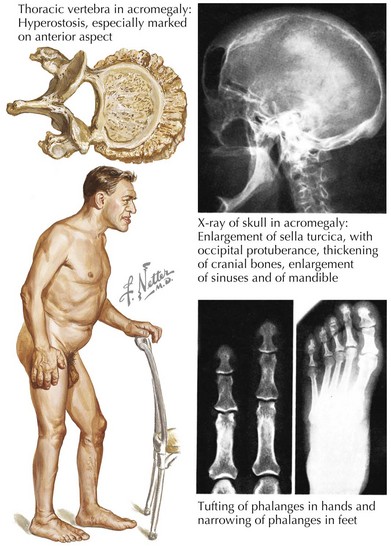
Sustained hypersecretion of GH by a pituitary tumor can lead to overgrowth of several tissues and to considerable cardiovascular changes (Fig. 61-1). Cardiovascular function is an important determinant of morbidity and mortality in untreated acromegaly. The most common comorbid cardiovascular condition accompanying acromegaly is hypertension, present in 50% of inadequately treated patients. Hypertension in acromegaly is usually mild but can be difficult to manage conventionally. Left ventricular (LV) mass can be significantly increased compared with that of normotensive patients. Curing the acromegalic condition is the most effective way to lower blood pressure (BP). A concentric ventricular hypertrophic cardiomyopathy unassociated with hypertension but associated with long-standing acromegaly develops in some patients and can result in both diastolic and systolic dysfunction. Cardiomegaly can be disproportionate to the changes in size that occur in other organs in severe acromegaly. The severity of cardiomyopathy correlates with the duration of exposure to high levels of GH. Diastolic dysfunction and hypertrophy develop first and are common in untreated patients. These changes are reversible with adequate treatment of GH excess. If left untreated, there is progression to systolic dysfunction, and heart failure and severe ventricular arrhythmias can occur. Histologic evaluation of the myocardium in patients with long-standing acromegaly shows interstitial fibrosis, lymphocytic infiltration, and sometimes necrosis.


Other changes in acromegaly can lead to secondary effects on the cardiovascular system. Some patients have sleep apnea that causes chronic recurrent hypoxemia, approximately 25% of patients have diabetes mellitus, and up to 40% of patients have hypertriglyceridemia. Premature mortality is increased in acromegaly, and cardiovascular diseases are the cause of death in 38% to 62% of patients. Normalizing GH and insulin-like growth factor 1 concentrations with conventional treatment restores normal life expectancy, preventing premature death resulting from cardiovascular disease.


Disorders of ADH Secretion


Unlike diseases of the anterior pituitary gland, the etiology of ADH deficiency is often hypothalamic lesions (in ~60% of patients). Most cases of ADH deficiency are acquired, and many result from attempts to remove the pituitary tumor surgically, damaging the pituitary stalk or the posterior pituitary. Severe ADH deficiency leads to polyuria, polydipsia, and, if untreated, vascular collapse. The most common hypothalamic causes are mass lesions, principally tumors of the hypothalamus, such as craniopharyngioma and dysgerminoma.


ADH is a potent pressor agent and stimulates direct vasoconstriction of blood vessels. This action is conferred at the level of the regional arterioles, and physiologic concentrations can induce this effect. Loss of ADH leads to a significant increase in serum osmolarity of greater than 295 mOsm/L, with inappropriately dilute urine of less than 300 mOsm/L. The diagnosis is established by detecting abnormally high serum osmolarity with low plasma vasopressin and low urinary osmolarity.


Administering vasopressin quickly reverses the changes in these parameters. Vasopressin acts on the kidney to increase free-water clearance. It also affects the brain to maintain central BP control; these brain actions are probably necessary for the maintenance of normal upright BP. The use of ADH antagonists illustrates the importance of endogenous arginine vasopressin for maintaining normal BP.


Thyroid Disorders


Hyperthyroidism


Hyperthyroidism causes some of the most impressive and sustained disarrangements of cardiovascular function related to endocrine abnormalities. Graves’ disease, the most common cause of hyperthyroidism, is triggered by an autoimmune process whereby thyroid antigens that are recognized as foreign stimulate the production of an autoantibody that stimulates the TSH receptor. The autoantibody directly binds to the TSH receptor on thyroid tissue and stimulates thyroid function. The effect of this stimulating antibody is unremitting and necessitates specific therapy to block thyroid hormone. The second most common cause of hyperthyroidism is a toxic multinodular goiter. This condition can account for 40% of the cases in patients over 60 years of age.


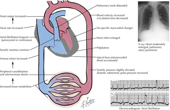
The symptoms of cardiac dysfunction that occur most commonly in thyrotoxicosis include fatigue, palpitations, dyspnea, heat intolerance, increased sweating, and weight loss. Tachycardia and palpitations occur in 80% to 90% of untreated patients (Fig. 61-2). Elderly patients in whom Graves’ disease develops may also experience heart failure. In this circumstance, the failing heart cannot meet metabolic requirements that are raised by increased thyroid hormone, resulting in overt congestive heart failure (CHF). Similarly, angina pectoris may be an important symptom in elderly patients with hyperthyroidism. Myocardial oxygen consumption can increase by as much as 70% in untreated hyperthyroidism. In the presence of fixed coronary lesions, blood flow may be inadequate to supply the increased metabolic need. In younger patients, thyrotoxicosis is associated with increased inotropic and chronotropic effects on the heart. Palpitations and occasionally atrial arrhythmias are the initial symptoms. Atrial fibrillation occurs in 33% to 47% of patients who are older than 60 years. Vascular resistance is decreased by peripheral vasodilation; the net effect is a marked increase in CO, which results in increased oxygen consumption. Peripheral edema is the most common symptom of overt heart failure in Graves’ disease, although dyspnea on exertion can also be prominent.


Physical findings typically include a hyperdynamic precordium, accentuated heart sounds, and a systolic murmur that can be heard over the precordium because of increased flow across the aortic valve. Mitral valve prolapse may also be present. Arrhythmias can range from sporadic premature beats to overt atrial fibrillation. Thyrotoxicosis is present in approximately 11% of patients with atrial fibrillation who are older than 60 years. Indeed, atrial fibrillation due to either hyper- or hypothyroidism is common enough that thyroid disease must be excluded at an early stage in the evaluation of this arrhythmia. ECG findings are nonspecific. Heart failure in younger patients is generally reversible with adequate treatment. Whether a distinct thyrotoxic cardiomyopathy exists is debated; however, extensive cardiac remodeling occurs in some patients. This may also be aggravated by long-standing tachyarrhythmias. In elderly patients in whom underlying cardiac abnormalities exist, heart failure can be severe and may trigger atrial fibrillation. Acceleration of angina pectoris can be dramatic in the elderly, and overt myocardial infarction can occur in these patients if left untreated.


The diagnosis is established by elevated serum thyroxine (T4) in the presence of a suppressed TSH concentration. Early in the disease, triiodothyronine (T3) is elevated, which is usually followed by a T4 elevation.


Initial treatment of Graves’ disease with antithyroid drugs blocks thyroid hormone synthesis. Treatment of the thyroid disease does not always restore normal sinus rhythm. If patients fail to undergo remission in a reasonable period on antithyroid drugs, or if they do not tolerate these medications, they are generally treated with radioactive iodine. In elderly patients with multiple cardiac complications, initial therapy with radioactive iodine may be indicated. While reversal of the thyrotoxic state generally restores cardiac abnormalities due to thyrotoxicosis to normal in younger patients, this is not always the case in elderly patients. Both sets of patients may benefit initially from therapy with β-blockers, which limits most effects of catecholamines on the cardiovascular system—effects that are accentuated in Graves’ disease. If a toxic multinodular goiter is present, the usual treatment is radioactive iodine.


An increasingly recognized, important cause of hyperthyroidism in cardiac patients is amiodarone-induced thyrotoxicosis (AIT). This usually occurs in patients during the first year of therapy. There are two different pathophysiologic mechanisms that induce hyperthyroidism. In the first (AIT type I), the iodine in aminodarone induces hyperthyroidism. These patients have a low radioactive iodine uptake, and color flow Doppler shows increased vascularity. They respond to potassium perchlorate and antithyroid drugs. Type II AIT patients have a distinctive thyroiditis induced by an amiodarone metabolite. These patients have a very low radioactive iodine uptake and absent vascularity. They respond to high-dose corticosteroid therapy.


Jun 12, 2016 | Posted by in CARDIOLOGY | Comments Off on Cardiovascular Manifestations of Endocrine Diseases

Full access? Get Clinical Tree

Get Clinical Tree app for offline access