(1)
Department of General surgery, Fuwai Hospital, Beijing, China
1.1 Nomenclature of Cardiac Anatomy
1.1.1 Right Atrium (RA)
The right atrium (RA) is the right upper chamber of the heart. It normally receives systemic venous drainage from the superior vena cava (SVC) and the inferior vena cava (IVC) (the two largest veins in the body collectively known as the venae cavae) and coronary venous drainage from the coronary sinus (Fig. 1.1). The RA can be divided into the body part, called the auricula, and the sinus part, called the sinus venarum. The auricula forms the front part of the RA and includes the right atrial appendage (RAA) and lateral wall, with numerous muscle bundles inside that form a network of hills and furrows, giving it a trabeculated surface. Located between the superior and inferior venae cavae, with an internodal tract passing through it, is a special muscle bundle called the crista terminalis. The sinus venarum is located just behind the crista terminalis and contains the orifices of the vena cava and coronary sinus.
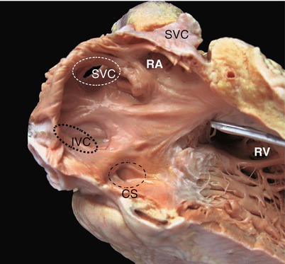

Fig. 1.1
The characteristics of the right ventricle (RV). The RV is a cardiac cavity directly connected with the SVC, the IVC, and the coronary sinus. CS coronary sinus, IVC inferior vena cava, RA right atrium, RV right ventricle, SVC superior vena cava
1.1.2 Right Atrial Appendage (RAA)
The RAA is a small conical, triangle-shaped muscular pouch. It is attached to the right atrium of the heart and often is short and blunt (Fig. 1.2).
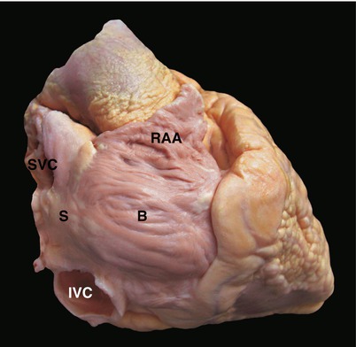

Fig. 1.2
The characteristics of the right atrial appendage (RAA). The RAA is short, blunt, wide-based, and triangle-shaped, with numerous muscle bundles inside that give it, like the RA, a trabeculated surface. B the body part of RA, RAA right atrial appendage, S the sinus venarum of RA, SVC superior vena cava, IVC inferior vena cava
1.1.3 Left Atrium (LA)
The term “left” when used for the left atrium (LA) is a morphological concept, rather than a simple direction. The LA is the cardiac chamber that receives drainage from the pulmonary veins through four ostia in its posterior wall. The LA has a smooth inner wall, whereas in the left atrial appendage (LAA), muscle bundles form a network of hills and furrows (Fig. 1.3). A crescent reductus can be seen in the interatrial septum, the wall of tissue separating the right and left atria, when looking through the LA, in contrast to the fossa ovalis, which can be seen when looking through the RA.
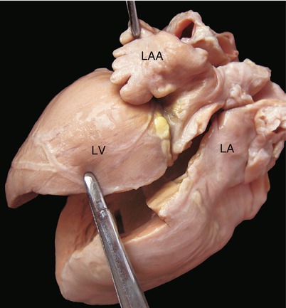

Fig. 1.3
Some muscle bundles form a network of hills and furrows in the LAA. This network is different from the inner wall of other parts of the LA
1.1.4 Left Atrial Appendage (LAA)
The left atrial appendage (LAA) is a small pouch located high in the LA. It pumps oxygenated blood from the lungs into the left ventricle. In contrast to the short and blunt RAA, the LAA has a long, narrow, and tubular shape and an obvious indentation; it usually is multilobed (Fig. 1.4).
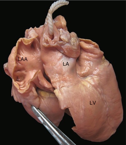

Fig. 1.4
In contrast to the RAA, the LAA has an obvious indentation that is just like the neck of the LAA. LAA left atrial appendage, LA left atrium, LV left ventricle
1.1.5 Right Ventricle (RV)
The right ventricle (RV) is a cardiac chamber located in the front side of the heart. It contains numerous muscle bundles and looks coarsely trabeculated. The term “right” is also a morphological concept. The morphological characteristic is that crista supraventricularis muscles widely separate the atrioventricular (tricuspid) valve from the semilunar (pulmonary) valve in the normal heart (Fig. 1.5). The inner surface of the RV consists of three parts, the orifice portion, the trabecular portion, and the outlet portion.
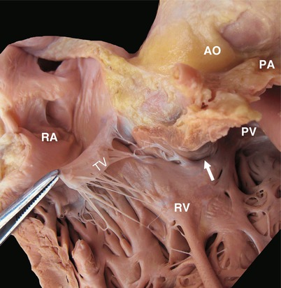

Fig. 1.5
The characteristics of the right ventricle (RV). The RV has numerous muscle bundles inside with coarse trabeculae. The atrioventricular or TV, with some chordae tendineae attached to the interventricular septum. The tricuspid valve is separated from the PF by the crista supraventricularis muscles. AO aorta, PV pulmonary valve, RA right atrium, RV right ventricle, TV tricuspid valve, SVC the white arrow show the muscle that separate pulmonary valve and tricuspid valve
The outlet portion of the RV is composed of muscles of the infundibulum. The muscle of the inferior border of the infundibular septum is called crista supraventricularis, or trabecula septomarginalis. It contains several different muscle bundles, the parietal band, the moderator band, and the septal band (Fig. 1.6). The septal band often extends apically to be continuous with the moderator band, which is an important trabeculation located among the septum, anterior papillary muscle of the tricuspid valve (TV), and the free wall. The crista supraventricularis is found between the TV and the pulmonary valve (PV).
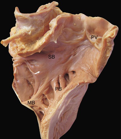

Fig. 1.6
The muscle bundles between the TV and the PV. MB moderator band, PB parietal band, PV pulmonary valve, SB septal band
1.1.6 Tricuspid Valve (TV)
The TV is the atrioventricular valve of the RV and has three leaflets: the anterior, posterior, and septal leaflets. The anterior leaflet is usually the largest one, with a strong anterior papillary muscle; the septal leaflet is usually smaller than the anterior leaflet but often larger than the posterior leaflet; it has a small chorda that mostly connects with the interventricular septum.
1.1.7 Left Ventricle (LV)
The left ventricle (LV) is a cardiac chamber that is divided into the orifice portion, the trabecular portion, and the outlet portion. The term “left” is also a morphological concept with regard to the LV. The mitral valve (MV) is the atrioventricular valve of the LV. In a normal heart, the MV is separated from the aortic valve (AV) by fibrous tissue instead of muscles.
1.1.8 Mitral Valve (MV)
The mitral valve (MV) is the atrioventricular valve of the LV and has two leaflets: anterior and posterior. These two leaflets connect with the anterior and posterior papillary muscles, respectively, by chordae. Normally, no chordae are attached to the interventricular septum.
1.1.9 The Posterior Cross of the Heart
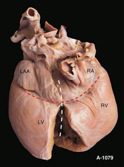
The posterior cross of the heart refers to the cross in the posterior aspect of the heart, located at the connection of the interatrial groove and the interventricular groove. The coronary sinus runs from the left to the right of the atrioventricular groove, forming the transverse line of the cross (Figs. 1.7 and 1.8). The posterior margins of the interatrial septum and the interventricular septum compose the upright line. The posterior margin of the interventricular septum is the same line as the posterior interventricular groove, with the posterior descending branch of the coronary artery passing through it.
 < div class='tao-gold-member'>


Only gold members can continue reading. Log In or Register to continue
Stay updated, free articles. Join our Telegram channel

Full access? Get Clinical Tree


