Figure 29.2 Images illustrate eccentric closing line of bicuspid aortic valve (a–d). LA, left atrium; LV, left ventricle; RA, right atrium; RV, right ventricle.
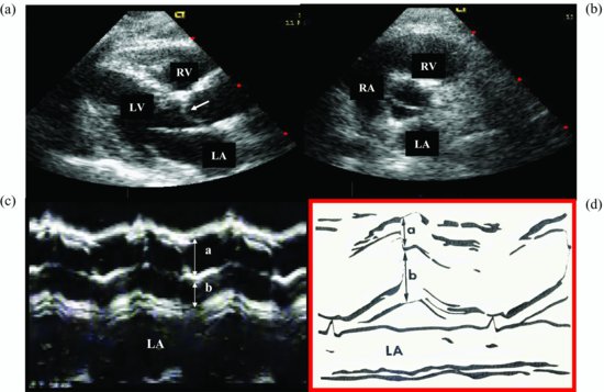
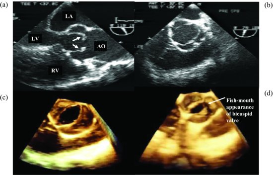
Figure 29.3 (a–d) Two-dimensional and three-dimensional images from parasternal window illustrate the different bicuspid aortic valves. AO, aorta; LA, left atrium; LV, left ventricle; RV, right ventricle.
Discussion
Stay updated, free articles. Join our Telegram channel

Full access? Get Clinical Tree


