Figure 36.1
(a) The patient undergoes general anesthesia and double-lumen intubation and is placed in a stretched lateral decubitus position, secured by a side-positioned cushion and elevated arm support. Bronchoscopy is performed if endobronchial lesions are suspected with symptoms such as hemoptysis and atelectasis; moreover, it is necessary to exclude endobronchial obstruction before attempting pleurodesis when the lung remains collapsed after thoracentesis. Surgical disinfection and sterile covering are extended from the axillary region down to the distal costal arch and from the mammary line backward to the vertebral column. A spacious sterile covering facilitates targeted localization of the surgical incisions in case of a loculated pleural effusion. Lung ventilation is terminated on the side of the operation. The first surgical incision is directed into the present pleural effusion to evacuate it and to obtain sufficient space for an initial exploration of the pleural surfaces. This incision is always made bluntly; digital intrathoracic exploration is performed to detect and release adhesions around the port site before the camera is inserted. (b) Further incisions are placed under video-optical control and are located at the furthest distance, either cranial in the third intercostal space if the first incision was located below the fifth intercostal space or into the diaphragmatic recesses if the initial approach was above the fifth intercostal space. For a diagnostic approach, two incisions usually are adequate; if a pulmonary wedge resection is required, a third incision may be appropriate

Figure 36.2
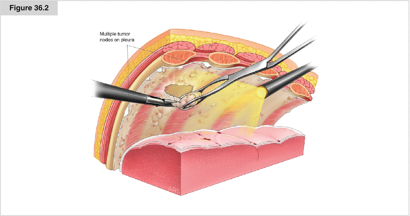
Gross inspection of the thoracic cavity reveals pleural adhesions, and the space for surgical intervention may be defined. The lung is mobilized and adhesions are dissolved by lung grasping forceps; a decortication may be done to achieve complete lung reexpansion. Subsequently, a complete video-optical investigation of the pleural surface (including the parietal and visceral pleura as well as the mediastinal and pericardial face and diaphragmatic surface, including the diaphragmatic recesses) is performed. The pleural surfaces (parietal and visceral) are examined for tumor growth, abnormal vascular inclusions, or thickening of the pleural layers. In cases of generalized pleural disease, up to three biopsy samples of the parietal pleura are extracted from different representative areas. If there is localized tumor growth, targeted biopsy samples are taken from the affected area. The extracted pleural specimens should include all layers of the parietal pleura to enable confident histopathologic diagnosis, including potential immunohistochemistry. If pleural mesothelioma is suspected and the patient is a candidate for extrapleural pneumonectomy (EPP) or complete parietal and visceral pleurectomy, tissue specimens should be taken from the diaphragm so as not to aggravate the extrapleural dissection during EPP or pleurectomy. Tissue specimens of the visceral pleura may be obtained best by pulmonary wedge resection. In cases of pleural asbestosis, tissue specimens should not be taken from the middle of a pleural plaque but from a border of the pleural plaques and should include the adjoining parietal pleura

Figure 36.3


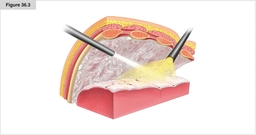
Controlled equilateral intraoperative ventilation confirms the dilatability of the lung and whether pleurodesis may be promising. If the lung is completely dilatable, 4 g of sterile talc is sprayed onto the pleural surfaces: the parietal and visceral pleura, mediastinal and pericardial face, diaphragma, and diaphragmatic recesses. At the end of this procedure, all surfaces are evenly covered with a thin layer of talc. Prior to talc insufflation, the pleural effusion has to be evacuated completely so the talc is not washed out. A chest tube is introduced, and controlled ventilation inflates the lung to contact the parietal and the visceral pleura. Postoperatively, the chest tube is connected to a continuous suction source for 24–48 h; the tube may be removed after radiologic proof of lung expansion at a regressive amount of pleural fluid below 150 mL/d
Stay updated, free articles. Join our Telegram channel

Full access? Get Clinical Tree


