Fig. 23.1
Cross-sectional anatomy of the infrapopliteal arterial system
The PT is easily approached through a longitudinal incision in the skin and deep fascia approximately 2–3 cm posterior to the tibia. The PT is immediately visible on the surface of the tibialis posterior muscle after incision of the deep fascia. The peroneal artery is deeper in the leg. It can be approached though a medial incision like the PT or can be exposed from a lateral approach after segmental resection of the fibula. The lateral approach to the peroneal is particularly appealing in the setting of significant soft tissue injury on the medial leg that could limit coverage of an arterial repair. The anterior tibial artery is exposed through a longitudinal incision in the skin and deep fascia approximately 2 cm lateral to the tibia. In most traumatic vascular injuries, the surgeon must make a decision whether to expose the injured vessels through traumatic skin lacerations or use more conventional incisions.
In making clinical decisions regarding the need for surgical intervention in lower leg trauma, it is critical to understand the redundancy of the blood supply to the foot. It may not always be necessary, and sometimes unwise, to reconstruct an isolated tibial artery injury in the absence of distal ischemia. In most cases, a single patent tibial artery is sufficient for distal perfusion of the foot and preservation of normal function. In the absence of hemorrhage, distal ischemia, or need for debridement of devitalized tissue, nonoperative management may be appropriate in selected patients. Non-emergent treatment of an intimal flap, arteriovenous fistula, or pseudoaneurysm can be undertaken electively.
23.4 Mechanism of Injury
Most penetrating injuries are due to gunshots, shotguns, and stabbing. Stab wounds usually result in isolated vascular injuries to both arteries and veins that can be treated by primary repair. In comparison to stab wounds, the extent of soft tissue damage, including vascular injury, is related both to the caliber and velocity of gunshots. High-velocity, military-style weapons may cause extensive damage well beyond the actual tract of the missile. In the assessment of vascular injury, the surgeon must be aware of the potential for vascular damage that is remote from the point of penetration. Shotgun wounds are notable for the inaccuracy of physical examination in detecting arterial injury, for more extensive soft tissue damage, and for the possibility of multiple vessel injuries.
Blunt arterial injuries may result from direct crushing, stretching, or laceration due to bony fragments. Blunt arterial injuries are often difficult to repair without vein graft interposition or bypass due to injury to a longer segment of arterial wall or delamination of the arterial wall with retraction of the intima. Both blunt and penetrating injuries that do not result in occlusion may result in pseudoaneurysm or arteriovenous fistula, which should be considered when planning follow-up surveillance. Regardless of the mechanism of injury, two principles govern operative repair: debride all nonviable vessels and complete the vascular repair without tension.
23.5 Clinical Assessment
The initial evaluation and resuscitation of trauma patients follows Advanced Trauma and Life Support (ATLS) protocols. Many patients with vascular injuries below the knee have other significant systemic injuries that must be evaluated and treated. This necessitates direct and clear communication between the vascular surgeon, the trauma surgeon, and other specialists. The vascular surgeon must be cognizant of systemic injuries in order to properly prioritize treatment of the injured extremity. It is particularly important to establish the presence of central nervous system injuries that might preclude anticoagulation. In addition, major vascular injuries in the chest, abdomen, or pelvis invariably take precedence over extremity vascular injuries.
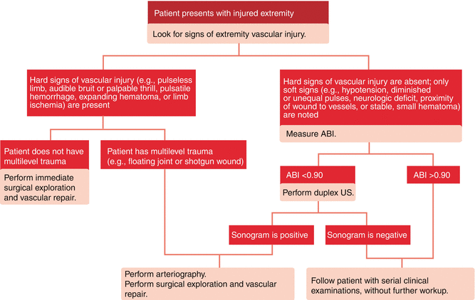
Following establishment of a stable airway, control of obvious bleeding, and initial resuscitation, a detailed examination of the extremities can be performed during the secondary survey (Fig. 23.2). For both blunt and penetrating injuries, the extremity examination is focused around four functional components: bone, nerve, soft tissue, and vessels. Injury to three of these four functional elements constitutes a “mangled extremity” [12]. The clinician should note the presence of limb deformity or instability suggesting an underling fracture. The extent of skin loss or soft tissue injury is noted. Neurological evaluation of the extremity is usually limited to basic motor and sensory function. The sciatic nerve supplies the muscles of the lower leg after it divides into the tibial nerve and the peroneal nerve. The tibial nerve supplies the muscles of the posterior compartment and the skin of the medial hind foot. Injury to the peroneal nerve results in the inability to dorsiflex the foot. The deep peroneal nerve courses through the anterior compartment and provides motor innervation to anterior compartment and sensation to the first and second toes. The muscles of the lateral compartment are supplied by the superficial peroneal nerve which also provides sensation to the skin of the anterolateral foot. The femoral nerve provides cutaneous branches to the anterior thigh, muscular branches to the pectineus, sartorius, the quadriceps femoris, and the vastus group of muscles.


Fig. 23.2
American College of Surgeons Lower Extremity Vascular Injury Algorithm
It is critical to perform a complete initial examination of the vascular system during the secondary survey in order to identify potentially lethal injuries and to repeat the exam at frequent intervals in order to detect significant changes. The presence, asymmetry, or absence of all peripheral pulses should be recorded in addition to the color and temperature of the extremity. Bruits and thrills in areas of suspected arterial injury may indicate the presence of significant stenosis, arteriovenous fistulae or pseudoaneurysm. The compartments of the affected extremity should be assessed subjectively for excessive pressure in comparison to the uninjured leg and measured objectively as necessary. Hematomas should be assessed for pulsatility or expansion. Distended superficial veins or excessive extremity edema may indicate proximal venous obstruction or the presence of arteriovenous communications.
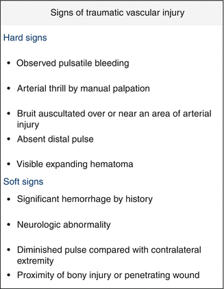
The evaluation of vascular injuries utilizes a group of clinical indicators commonly referred to as “hard” and “soft” signs (Fig. 23.3). The hard signs of vascular injury include observed pulsatile bleeding; absent distal pulse; expanding hematoma; audible bruit; palpable thrill; or a cold, pale, or mottled extremity. The sensitivity of any hard sign is nearly 100 % in the detection of significant vascular injury [13]. In the absence of other higher-priority, life-threatening injuries, the presence of a hard sign of vascular injury typically indicates the need for emergency surgical treatment. In a patient with obvious extremity bleeding, the patient should be taken directly to the operating room for immediate exploration. In a patient with limb ischemia, expeditious vascular imaging, optimally performed in the operating room, prior to surgical exploration may be preferable to blind surgical exposure, particularly in patients with multiple sites of suspected injury. Recent experimental evidence suggests that the duration of limb ischemia is related to the magnitude of detrimental systemic effects following reperfusion, further emphasizing the need to avoid excessive delay in attempts to obtain preoperative imaging [14]. As previously discussed, in a patient without clinical signs of overt ischemia, the absence of an isolated pedal pulse may warrant nonoperative management particularly in the setting of other significant nonvascular injuries. Elective vascular imaging with color flow Doppler and CT-, MR-, or catheter angiography to rule out significant intimal flap, pseudoaneurysm, or arteriovenous fistula is appropriate in stable patients without ischemia if a nonoperative approach is elected.


Fig. 23.3
Clinical indicators of vascular injury
The “soft” signs of vascular injury include a history of pulsatile bleeding, diminished pulse compared to the contralateral uninjured extremity, proximity of bony fragment or penetrating wounds adjacent to arterial structures, and isolated neurologic deficit of the injured extremity. The specificity of “soft signs” for the detection of significant arterial injury is less than 5 %.
As an adjunct to the physical examination, the ankle-brachial index (ABI) may aid assessing the presence and severity of vascular injury. The ABI is the ratio of the highest blood pressure detected by continuous wave Doppler at the level of the ankle of the injured lower extremity divided by the highest blood pressure in an uninjured upper extremity. Both upper and lower extremity blood pressure measurements are performed using the Doppler rather than a stethoscope. The ABI may be performed at the bedside with only a manual blood pressure cuff and a continuous wave Doppler and does not require advanced imaging equipment or a certified vascular technologist.
The accuracy of Doppler-derived ABI in patients with lower extremity vascular injuries has been reported with both penetrating and blunt lower extremity trauma. Lynch and Johansen examined the sensitivity and specificity of ABI measurements in trauma patients with suspected lower extremity vascular injury [15]. In this series of 100 consecutive injured limbs that were evaluated by Doppler-derived ABI and contrast angiography, an ABI of less than 0.9 was 87 % sensitive and 97 % specific for the detection arterial injury. From these data the authors suggested that in the absence of hard signs of arterial injury, an ABI is an acceptable alternative to angiography particularly if there is an anticipated period of continued observation [16].
The use of ABI has also been applied to the evaluation of patients with blunt vascular injuries of the lower extremity associated with fractures and dislocations. A controlled trial that included 75 consecutive patients with knee dislocations that were believed to be at high risk for vascular injury was performed to determine the diagnostic accuracy of ABI [17]. The negative predictive value of a Doppler-derived ABI greater than 0.9 was 100 %. Of the 30 % of patients with an ABI less than 0.9, 70 % had an injury that was diagnosed by angiography, 50 % of which required repair. This study again demonstrates the value of a simple, noninvasive diagnostic test that provides important objective information that can be performed rapidly at the bedside [17].
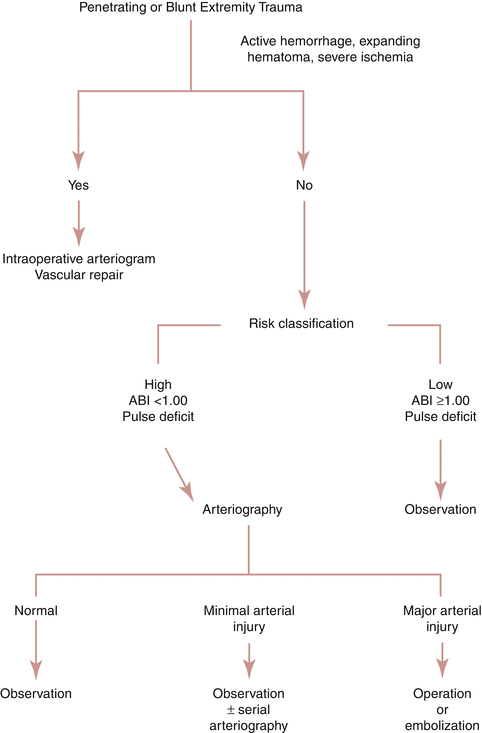
Despite the obvious advantages of Doppler-derived ABI in the trauma setting, there are several situations and patient characteristics that may render the ABI inaccurate. Injuries to non-axial lower extremity arteries such as the profunda femoris or the peroneal artery may exist despite a normal ABI. Also, a nonobstructive intimal flap, an arteriovenous fistula, or a pseudoaneurysm may not be associated with a decrease in the ABI. In these situations a heightened clinical suspicion based on mechanism of injury and anatomic location of injury is instrumental in pursuing further diagnostic testing (Fig. 23.4).


Fig. 23.4
Diagnostic algorithm for lower extremity vascular injury
23.6 Diagnostic Imaging
23.6.1 Selective Contrast Angiography
Mandatory surgical exploration of “proximity” extremity wounds was largely abandoned in the 1970s in favor of less invasive vascular imaging [18]. Angiography has a sensitivity of >95 % and specificity of >90 % in detecting arterial injury [19]. In addition, diagnostic angiography- and combined with catheter-based intervention has emerged as a valuable treatment option in certain situations such as active hemorrhage, pseudoaneurysm, occlusion, and AV fistula in both blunt and penetrating injuries. On the other hand, angiography is invasive and has many well-recognized complications including puncture site complications, contrast nephropathy, allergic reactions, and remote vessel injury.
23.6.2 Computed Tomographic Angiography
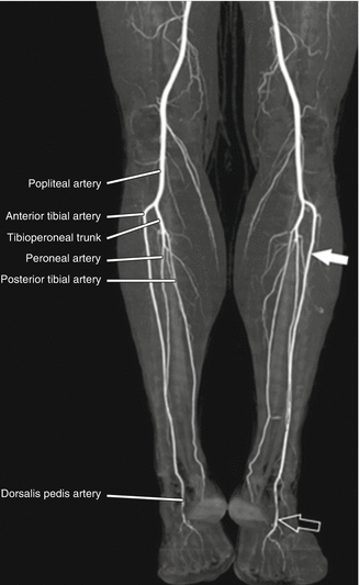
Multi-slice helical computed tomographic angiography (CTA) has been increasingly used in the evaluation of lower extremity vascular injuries in major trauma centers [20]. Major improvements in CT technology and its widespread availability have resulted in considerable reduction in the use of catheter-based angiography in the evaluation of extremity trauma (Fig. 23.5). CTA provides a rapid, noninvasive diagnostic tool that provides three-dimensional information regarding arterial injury as well as other soft tissue and bone injuries in the same anatomic region. The sensitivity and specificity of CTA for extremity vascular trauma has been demonstrated to be in excess of 95 % for both penetrating and blunt injuries to large proximal vessels [21].


Fig. 23.5
CTA imaging of normal infrapopliteal arteries. The solid arrow indicates a dominant anterior tibial artery and the open arrow shows the dorsalis pedis artery at the ankle
Despite widespread use in the evaluation of trauma patients, there are a number of disadvantages associated with CTA that must be considered in evaluating potential injuries to the small arteries of the lower leg. Errors in timing and dose of the contrast bolus may result in inadequate visualization of the tibial vessels. Spasm associated with shock and hypothermia may further limit the adequate visualization of the distal vasculature. In addition, care must be taken to provide continuous monitoring of ventilation and hemodynamics during transport and transfer since the patient may be relatively inaccessible in the event of sudden clinical deterioration. Despite these disadvantages, CTA continues to gain widespread use in the evaluation of vascular extremity trauma.
23.6.3 Duplex Ultrasonography
Duplex ultrasonography (DUS) combines color flow Doppler ultrasound with conventional ultrasound to render high resolution imaging of vascular structures. The combination of the two modalities allows for the identification and imaging of specific arterial and venous structures. Duplex ultrasonography has been shown to be a reliable method of diagnosis in patients with potential vascular injuries and has been incorporated into a variety of trauma protocols. Sensitivity and specificity of duplex ultrasound in the evaluation of extremity vascular injury have been reported as high as 95 % with overall accuracy between 96 and 98 % [22, 23].
There are several advantages that make DUS an attractive option for evaluating vascular injury. It is noninvasive and painless, can be performed in the trauma resuscitation area, and is easily repeated. It provides information regarding the character of arterial flow, identification of active hemorrhage, dissection, pseudoaneurysm, and AV fistula. In addition, duplex ultrasound has the benefit of allowing interrogation of the venous system which frequently harbors occult injury [24].
Despite the aforementioned advantages of DUS, there are several important disadvantages and clinical situations where it may not be appropriate. DUS is highly operator dependent and may not be immediately available depending on local resources. It may not be possible to perform a complete arterial evaluation of an injured lower extremity due to overlying bandages or external fixation. Lastly, the inability of the patient to cooperate with the study due to pain, open wounds, or impaired state may limit the effectiveness of DUS. If any of these situations negatively impacts the performance of the DUS, then alternative imaging will be required.
23.7 Initial Management
Management can be organized by indication: hemorrhage, ischemia, and proximity. For patients with hemorrhage from extremity vascular injury, immediate surgical exploration is indicated. For patients with other hard signs of vascular injury, expeditious transfer to an operating room for image-directed surgical repair is appropriate. In the patient with soft signs of arterial injury or an abnormal ABI <0.9, additional imaging to identify arterial injury is recommended. In the setting of ischemia, despite the widely published recommendation that blood flow must be restored within 6 h of injury to avoid permanent ischemic injury, there is no absolute time limit beyond which vascular repair is absolutely contraindicated [25]. The degree of clinical ischemia is dependent on a number of factors in addition to the absolute duration of warm ischemia including the adequacy of collateral circulation, the presence of simultaneous venous obstruction, and overall hemodynamic stability. The vascular surgeon must take these factors into consideration in determining the optimal timing of surgical intervention.
Special discussion of the patient with suspected vascular injury in the association with fracture or dislocation is warranted. In the patient with a displaced fracture, the pulse and ABI should be determined both before and after orthopedic manipulation. If orthopedic manipulation preserves or restores a normal palpable pulse and ABI, additional imaging is probably unnecessary. If orthopedic manipulation is not accompanied by preservation or prompt restoration of normal distal perfusion, then additional vascular management is based on the severity and duration of ischemia. Direct communication between the orthopedic and vascular surgeon is critical in this setting to determine the optimal sequence of operative events. In most cases in the absence of profound ischemia (e.g., audible but abnormal Doppler signal at the foot), the orthopedic procedure should be completed prior to vascular reconstruction. If external fixation is selected, the orthopedic and vascular teams must coordinate the location of the orthopedic hardware to allow surgical exposure of the vascular injury. This usually entails placement of orthopedic hardware along the lateral aspect of the lower leg to allow medial exposure of the tibial vessels. Fracture fixation prior to vascular repair has the benefit of providing superior stability of the vascular repair and assures that the vascular reconstruction will not be compromised when the fracture is distracted and realigned.
Early restoration of blood flow prior to orthopedic fixation is appropriate in patients with profound ischemia (absence of Doppler signal and significant motor/sensory deficit) particularly if the period of warm ischemia will exceed 4–6 h. In selected cases, an intraluminal shunt can be used to temporarily restore arterial flow during orthopedic manipulation. The technique of emergency vascular shunting is relatively straightforward. We prefer to use an outlying (30 cm) Sundt shunt that is designed with conical shoulders at each end to prevent dislodgement. We prefer to secure the shunt in place with umbilical tape tourniquets or large “vessel loops” which are nearly universally available. Flow through the shunt should be demonstrated with Doppler. Venous shunts are usually unnecessary and occasionally meddlesome. In general, we recommend formal vascular repair immediately after the orthopedic procedure is completed although prolonged use of a temporary shunt for more than 24 h has been reported.
23.8 Operative Preparation
The operating team should be prepared for unanticipated events. Special instruments for arterial repair including fluoroscopy should be available. The patient should be positioned on a table that is suitable for fluoroscopic imaging. The room should be arranged to accommodate additional equipment including cell saver and imaging equipment. Preoperative antibiotics with gram-positive coverage should be administered prior to skin incision. Additional gram-negative coverage should be administered if there are associated fractures. The injured lower extremity should be draped and prepped widely taking into consideration a proximal point that may be required to obtain arterial control or provide inflow for bypass. The contralateral leg should also be prepped into the field in the event that the contralateral greater saphenous vein (GSV) or deep femoral vein is required for arterial reconstruction.
23.8.1 Surgical Treatment
The initial step in surgical management is to obtain proximal control prior to exposure of the arterial injury. In general, proximal control should be established as close to the injury as possible without risking hemorrhage due to inadvertent entry into the injury site. Initial proximal control at the level of the common femoral artery will usually prevent life-threatening exsanguination from an extremity vascular injury, but will not provide sufficient hemostasis to allow completion of the arterial repair. The above-knee popliteal artery is exposed through a medial incision and retraction of the sartorius and vastus medialis muscles. For more distal injuries, proximal control of the below-knee popliteal artery is appropriate. Exposure is obtained through an 8–10 cm incision located a fingerbreadth behind the tibia followed by posterior retraction of the medial head of the gastrocnemius. The popliteal vein should be retracted toward the table to expose the artery and to avoid injury to the tibial nerve. Temporary proximal control using an extremity tourniquet is an acceptable alternative. The tourniquet should be deflated when conventional vascular control is obtained.
In the absence of a contraindication such as intracranial hemorrhage, extensive soft tissue injury, or intra-abdominal solid organ fracture, we prefer to administer systemic intravenous heparin (50 units/kg) during the period of vascular occlusion. Local instillation of dilute heparinized saline (1 unit/cc) into the inflow and outflow vessels is a satisfactory alternative if the risk of systemic anticoagulation is thought to be excessive. In either case, the surgeon should be prepared to use a Fogarty embolectomy catheter at the completion of the procedure to assure complete clearance of thrombus from the runoff.
Once proximal control is obtained, the area of injury can be directly exposed. Care should be taken to avoid overly aggressive dissection to prevent iatrogenic vascular injury. The following principles apply to all vascular injuries below the knee:
1.
Vascular repairs should be tension-free. With the exception of the popliteal artery, primary repair is not usually possible for injuries that require resection of even very short vessel segments. Interposition vein graft should be considered if there is any concern that the reconstruction will be under excessive tension.
2.


Debride all devitalized tissue. This is particularly important for arteries that have been crushed or injured by high-velocity bullets. To the degree possible, it is best to resect all injured artery until normal endothelium is visualized.
Stay updated, free articles. Join our Telegram channel

Full access? Get Clinical Tree


