Balloon Catheter Embolectomy for Macroembolization in the Extremities


Balloon Catheter Embolectomy for Macroembolization in the Extremities


Dawn M. Coleman


Acute lower extremity ischemia remains a common vascular disease with considerable associated morbidity, limb loss, and mortality. Both arterial embolism and thrombosis are recognized causes of acute limb ischemia. It is imperative that a distinction between the two be made preoperatively because this guides the appropriate approach to therapy.


Surgical embolectomy remains the treatment of choice in managing most cases of acute embolic limb ischemia because it offers more rapid restoration of blood flow in comparison to catheter-directed thrombolysis. Time often becomes critical when considering the near absence of well-developed arterial collaterals in acute arterial macroembolization, when compared to in-situ thrombosis of chronic vascular occlusive disease. Additionally, in settings of acute limb ischemia, embolectomy is highly protective against amputation and, potentially, mortality.


Catheter-based intervention, first introduced by Fogarty in 1963, simplified the surgical technique of embolectomy. Although the technical details have not evolved dramatically since that time, a number of additional catheter options and adjuncts have been developed to effectively manage residual thrombus and underlying chronic disease.


Preoperative Diagnosis and Management


Patients who come to the hospital with acute limb ischemia require a thorough history and physical examination. Embolism should be suspected with a history of atherosclerotic or valvular heart disease, proximal arterial aneurysm, cardiac arrhythmia, and preceding trauma (iatrogenic or otherwise). Patients with embolic occlusion complain of acute symptom onset. They typically do not support a history of claudication or rest pain and have a normal contralateral pulse. The exception to this is the patient who comes to the hospital with aortic saddle embolism, paraplegia, and loss of bilateral femoral pulses. Symptom duration is important, and one should clinically categorize and document the ischemic limb as salvageable or not based on baseline motor, sensory, and Doppler examination. Physical examination should indicate the level of arterial occlusion, because signs of ischemia are often most pronounced one joint distal to the level of occlusion.


Upon arrival at the hospital, patients with acute limb ischemia should receive heparin to prevent thrombus propagation and maintain collateral patency, reducing the extent of ischemic injury. Additionally, the documented antiinflammatory effects of heparin protect some against ischemia-reperfusion injury. Appropriate and indicated medical resuscitation should be achieved if possible preoperatively (i.e., correcting any malignant arrhythmia, acidosis, or metabolic derangement). Noninvasive duplex imaging can efficiently aid in diagnosis and operative planning. Arteriography should be considered in patients with Rutherford class IIA disease, especially if the diagnosis of embolism is not certain.


Although femoral and brachial embolectomy may be performed with local anesthesia, distal exposures and certain adjunctive measures can require general anesthesia. Any invasive intraoperative monitoring (i.e., central venous catheterization, pulmonary artery catheterization, transesophageal echocardiogram) should be considered on a case-by-case basis as determined by medical comorbidities. Arterial line placement facilitates intraoperative surveillance of the activated clotting time and allows close blood pressure monitoring, which can become labile at the time of reperfusion.


Operative Technique


The lower abdomen, bilateral groins, and circumferential affected limb should be surgically prepped and draped in a standard fashion when intervening for lower extremity embolism. Baseline activated clotting time should be assessed and heparin should be dosed to maintain a level 250 seconds or longer for the duration of the procedure. A longitudinal incision made over the course of the femoral artery bifurcation is favored to facilitate cannulation of the common, superficial, and profunda femoral arteries and to allow the clot to be evacuated from the entire leg. Potts vessel loops around each vessel typically suffice for vascular control in the absence of a palpable pulse. Conventional arterial clamping risks fracture of the embolus, although vascular clamps may be used later for definitive control following successful thromboembolectomy. The balloon catheter size should be selected thoughtfully because a catheter too small might not achieve complete thromboembolectomy and a catheter too large can risk an operative vessel injury with thrombosis or an arterial disruption with formation of a pseudoaneurysm.


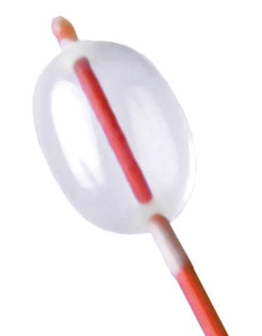
A transverse arteriotomy of a normal size and a nondiseased common femoral artery is favored, although a longitudinal arteriotomy should be considered if there is suspicion for underlying chronic occlusive disease that might require adjunct endarterectomy with patch angioplasty or femoral–distal bypass. Before the arteriotomy, the surgeon should confirm the embolectomy balloon is concentric during inflation with saline (Figure 1). Attempts at restoring inflow should start with the retrograde passage of a 5-Fr catheter through the common femoral artery. The catheter should be advanced to the level of the iliac bifurcation (approximately 50 cm), the balloon should be inflated with saline (not air), and the catheter should be withdrawn without releasing tension on the catheter. The person who is controlling catheter removal should control the balloon inflation. This maneuver should be repeated until the balloon returns clean of further thrombus and there is robust arterial inflow. In cases of an aortic saddle embolus, it is imperative that manual compression of the contralateral groin and femoral artery be maintained during balloon withdrawal to decrease the risk of distal thromboembolism in the opposite lower extremity. The retrieved embolic material should be sent for both culture and pathology assessment if there is any concern for infectious endocarditis or atrial myxoma.


Stay updated, free articles. Join our Telegram channel

Aug 25, 2016 | Posted by in CARDIOLOGY | Comments Off on Balloon Catheter Embolectomy for Macroembolization in the Extremities

Full access? Get Clinical Tree

Get Clinical Tree app for offline access