Axillofemoral Bypass


Axillofemoral Bypass


Joseph R. Schneider


Axillofemoral bypass (AxFB), a bypass graft from one axillary artery to one or both femoral arteries, was first performed in the early 1960s in patients at high cardiopulmonary risk for surgery as an alternative to the more direct reconstructive operation, an aortofemoral bypass (AFB). Initially, AxFBs were placed in patients with bilateral iliac artery occlusive disease, but the operation was quickly extended to reconstruction in patients with aortic infection (mycotic aneurysm), infected aortic prostheses, and other intraabdominal infections or otherwise hostile abdomens. At first the operation was most likely viewed as a technique to be used for limb salvage only in patients with critical limb ischemia or in those whose normal axial flow was expected to be interrupted by aortic ligation or removal of an aortic prosthesis. As the operation became familiar to surgeons, it was in some cases extended to patients with ischemic claudication.


A primary area of controversy in the 1970s to 1990s was whether this operation is in fact appropriate for patients with claudication. This question is complex. Claudicants tend to have less systemic atherosclerotic and other serious disease, they tend to live longer and enjoy better graft patency, and series dominated by claudicants tend to have more favorable results as measured by long-term graft patency and patient survival.


On the other hand, series composed primarily of patients with chronic critical limb ischemia and certainly those with a large fraction of patients with aortic and aortic prosthetic infection are associated with less favorable patient survival. Patients with critical limb ischemia have less favorable graft patency. It is the author’s perspective that the claudication question has never been settled, at first because patient selection was clearly so different among reported case series and more recently because endovascular techniques can be effectively applied in a significant fraction of patients who would have been considered for an AxFB in the past. Even aortic infection or an infected aortic prosthesis is a less secure indication for an AxFB because many of these patients have been successfully managed with in situ aortic graft replacement using antibiotic-treated prosthetic grafts, autologous superficial femoral veins, or arterial homografts.


General Principles and Patient Selection


Successful AxFB depends on the ability of one axillosubclavian artery to supply adequate blood for the arm and one or both legs. In general, the evidence is that in the absence of an occlusive process in the axillosubclavian arterial system, this concept is sound and the operation will provide adequate resting perfusion to both legs without causing harm to the arm. However, some patients have overt or occult disease in the inflow to the donor axillary artery, most often at the origin of the brachiocephalic or subclavian artery. One group has recommended routine arch and axillosubclavian angiography before performing an AxFB. We have not found this to be necessary in practice and are satisfied with demonstration of a normal triphasic Doppler waveform in the brachial artery and no more than a 10 mm Hg lower systolic blood pressure in the proposed donor-side arm compared to the contralateral arm.


AxFB is most often considered for high-risk patients with aortoiliac arterial occlusive disease who cannot be treated by endovascular techniques or with bypass from the contralateral femoral or other, closer donor artery. Endovascular techniques for arterial occlusive disease have advanced dramatically since the 1990s, and in most patients and most practices these endovascular techniques would now be primary treatment for these patients, if feasible. The concept of prohibitive risk, usually a result of advanced cardiopulmonary disease, is not well defined. Furthermore it is likely that many patients who would in the past have been considered to be at prohibitive risk as a result of cardiopulmonary or other systemic disease would now be viewed as average risk in vascular surgery practice.


However, unreconstructed coronary artery disease with recent myocardial infarction, angina, or abnormal stress testing would certainly be considered high risk for AFB. Compromised pulmonary function with FEV1.0 less than 1 L would also be considered very high risk for AFB. With respect to hostile abdomen, this would generally include problems of intraabdominal infection such as peritonitis or abscess, the presence of an intestinal or urinary stoma, or previous infection or prior surgery with adhesions, and these are potential indications for an AxFB. It is likely that the majority of patients with infrarenal aortic infection or infected aortic prosthetic grafts are still managed with aortic débridement and ligation with excision of the infected graft and AxFB, although there is no way to determine this with certainty, and there are some alternatives in such cases.


Preoperative Evaluation


In addition to blood pressure check and Doppler interrogation of the proposed donor-side brachial artery, a lower extremity arterial segmental Doppler waveform and pressure study with toe pressures should be obtained. This study helps confirm the diagnosis and calibrate the severity and also serves as a baseline to allow assessment of the results of any intervention. An angiogram, most often either computed tomography (CT) angiogram or conventional transfemoral or transbrachial catheter angiogram, is required for planning the intervention. In most cases an angiogram will have been performed before an AxFB is even considered. General medical evaluation, often including some sort of cardiac stress testing, is performed to better assess risk and guide the choice of intervention.


Technique


The operation is performed with the patient supine and the arm on the donor side abducted to 90 degrees, nearly always with general anesthesia. Towels, gel pads, intravenous fluid bags wrapped in towels, or other suitable padding is placed between the patient and the operating table on the side of the axillofemoral component to elevate the flank and lower chest. The patient is prepped from the neck to the lower anterior thighs including the intervening abdomen, ipsilateral flank, and chest including the sternum to allow entry to the abdomen and ipsilateral chest or a sternotomy (although none of these has ever been required in the author’s practice). Perioperative prophylactic antibiotics are administered and continued for 24 hours. Specific antibiotic coverage is administered as necessary in cases of an established remote infection when surgery cannot be delayed in an attempt to clear the infection.


The donor axillary artery is exposed using a transverse incision inferior to the middle of the ipsilateral clavicle, splitting of the pectoralis major muscle fibers, and division of the deep fascia. Exposure and control of the axillary artery is in most cases facilitated by ligation and division of at least one large crossing vein. Care must be taken to avoid injury to the adjacent axillosubclavian vein and brachial plexus elements. The axillary artery is less robust than the more familiar femoral artery and must be treated with great care to avoid injury. The author does not divide the pectoralis minor muscle as some have recommended. A critical technical point is to keep the graft anastomosis as medial as possible on the axillary artery to reduce the tendency for later arm abduction to disrupt the anastomosis. Leaving the pectoralis minor intact forces the surgeon to keep the anastomosis medial. One or both femoral arteries are exposed through standard groin incisions. A longitudinal groin incision is used most often because it allows more flexibility depending on findings at inspection of the target arteries.


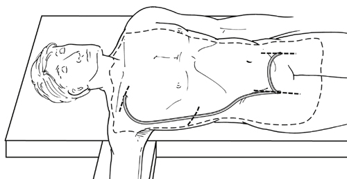
The axillofemoral graft is tunneled from the axillary artery exposure to the femoral exposure, which is nearly always ipsilateral (Figure 1). Circumstances requiring bypass from one axillary artery to a single contralateral femoral target are rare. The author’s team’s practice is to tunnel the graft posterior to the pectoralis minor muscle, but the graft may be tunneled anterior to the pectoralis minor, or the pectoralis minor may be divided, and there is no evidence that this affects outcome. The graft should be tunneled in the subcutaneous tissue in the midaxillary line to prevent kinking during subsequent patient torsoflexion and compression/angulation of the graft by the costal margin, which tends to be more prominent anteriorly. Classic descriptions of the operation have included an intermediate incision in the flank to facilitate tunneling, but we prefer to use a long enough tubular tunneler that allows tunneling without any intermediate incision.


Stay updated, free articles. Join our Telegram channel

Aug 25, 2016 | Posted by in CARDIOLOGY | Comments Off on Axillofemoral Bypass

Full access? Get Clinical Tree

Get Clinical Tree app for offline access