Atypical Carcinoid
Presentation
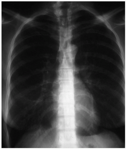
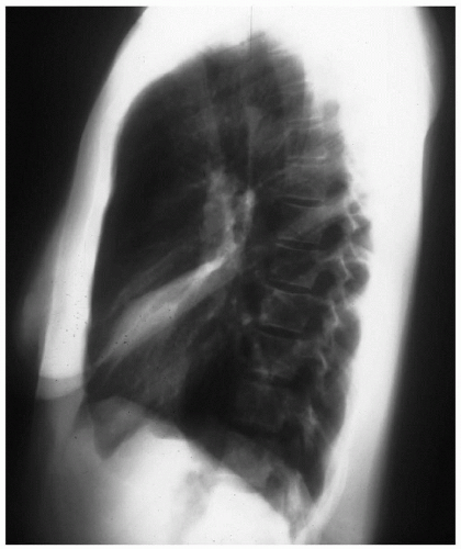
A 14-year-old boy presents to the emergency department complaining of a cough and dyspnea for the past 2 weeks. The patient does not report elevated temperature, chills, or sweats. An evaluation performed in the emergency department includes a chest x-ray, which is interpreted as showing right lung pneumonia. Oral antibiotics are administered, and the patient is discharged home. Over the next few weeks, the patient’s symptoms fail to improve, and a repeat chest x-ray is performed. Physical examination is significant for diminished breath sounds and wheezing at the right lung base.
Case Continued
Based on the chest x-rays, a different course of antibiotics is instituted with some mild improvement. A Mantoux purified protein derivative (PPD) skin test is obtained but is nonreactive. The radiographic abnormality does not resolve after the second course of antibiotics, and chest computed tomography (CT) scans are obtained to delineate the findings on chest x-rays.
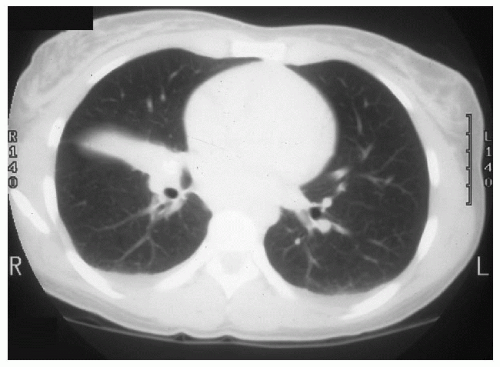
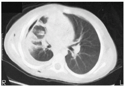
▪ CT Scans
CT Scan Report
The scan demonstrates a right hilar density measuring 3 cm with no evidence of calcification. No other enlarged hilar or mediastinal nodes are identified. The lung windows show segmental atelectasis of the right middle lobe. On additional images, right hilar adenopathy with segmental atelectasis of the right middle lobe is noted. These findings are consistent with right middle lobe syndrome.
Discussion
Right middle lobe syndrome is a bronchial compressive disease assumed caused by extrinsic compression of the airway by peribronchial adenopathy. Symptoms typically include productive cough, hemoptysis, recurrent fever, and chest pain. Chest x-ray studies demonstrate a collapsed middle lobe, with or without associated adenopathy. Because other causes of bronchial obstruction exist, a definitive diagnosis needs to be established.
Recommendation
Flexible bronchoscopy.
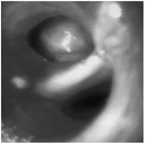
▪ Flexible Bronchoscopy Photograph
Bronchoscopy Findings
At bronchoscopy, a smooth, red-tan tumor is identified obstructing the right middle lobe bronchus. A biopsy is performed.
Stay updated, free articles. Join our Telegram channel

Full access? Get Clinical Tree


