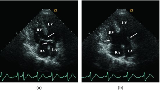
The apical 4-chamber echocardiographic images show: There is a large hole in the center of the heart. It’s located where the septum between the atrial chambers joins the wall between the ventricular chambers (arrows) (A). The common atrioventricular defect is seen spanning the large defect in the atrioventricular septum; the arrows point the leaflets of common valve (Figure 46.2).
Figure 46.2 The apical 4-chamber views show: There is a large hole in the center of the heart. It’s located where the septum between the atrial chambers joins the wall between the ventricular chambers (arrows) (A). The common atrioventricular defect is seen spanning the large defect in the atrioventricular septum; the arrows point the leaflets of common valve (B). LA, left atrium; LV, left ventricle; RA, right atrium; RV, right ventricle.
The left to right shunt and right side regurgitation are detected by color Doppler from parasternal long axis and apical 4-chamber views (Videoclip 46.1).
Discussion
Stay updated, free articles. Join our Telegram channel

Full access? Get Clinical Tree


