Aspergilloma
Presentation
You are called by the emergency department to evaluate a 45-year-old Vietnamese man with hemoptysis. He reports having streaks of blood in his sputum for the past few weeks but has now coughed up about 30 mL of blood. He is hemodynamically stable without respiratory distress.
Past medical history is significant for tuberculosis diagnosed 2 years ago and treated with isoniazid, rifampin, pyrazinamide, and ethambutol for a period of 6 months.
Review of systems is significant for recent fevers and cough.
Differential Diagnosis
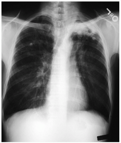
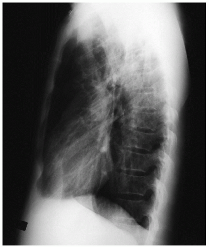
The opacity seen on the chest x-rays could represent an infectious or neoplastic process. This patient’s complaints of fever, cough, and hemoptysis are consistent with either diagnosis. However, given the history of previous treatment for tuberculosis, a superinfection or reactivation tuberculosis is the more likely diagnosis.
Discussion
Patients with chronic lung diseases are prone to developing fungal infections. In the case of tuberculosis, aspergillus is the most common organism. Hemoptysis is frequently present and can occur as life-threatening hemorrhage. The underlying tuberculosis is typically active in 20% to 50% of cases.
Recommendation
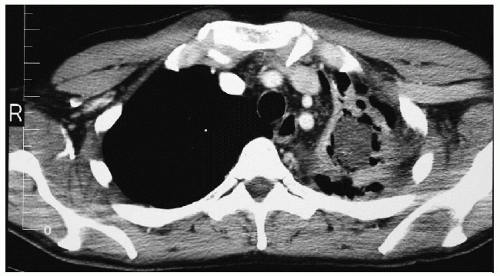
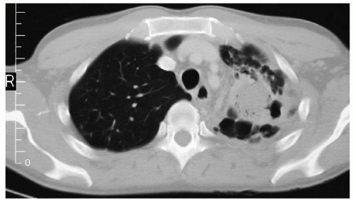
Computed tomography (CT) scans of the chest to delineate the left upper lung field process.
▪ CT Scans


