Arteriomegaly and Aneurysmosis


Arteriomegaly and Aneurysmosis


Bruce Torrance, Malachi Sheahan and Larry Hollier


Arteriomegaly is defined as a diffuse ectasia involving multiple arterial segments including the aorta, iliofemoral, and femoropopliteal vessels. It is characterized by significant tortuosity, elongation, and luminal irregularities often resulting in diminished blood flow. It was originally described in 1971 by Thomas, with reference given to two cases described by Leriche in 1942 and 1943. These patients were noted to have “extraordinary elongation and dilatation of the pelvic and common femoral arteries.” Leriche termed this condition “arteria magna et dolicho.”


In itself alone, arteriomegaly is considered to be a benign condition; however, aneurysmal involvement can and commonly does affect these arteries. It can be quite extensive. This process is distinct from general atherosclerotic aneurysms and is termed aneurysmosis or arteriomegaly with aneurysms. An increased morbidity and mortality is seen with aneurysmosis, and given the extent of disease, it can necessitate extensive and atypical surgical treatment.


Etiology


The cause of arteriomegaly and aneurysmosis is not well understood. Histological studies of arteries of patients with this disease have shown changes consistent with a connective tissue disease, specifically degradation of elastin in the medial layer, as well as arteriosclerosis. Although no distinct genetic component has been identified to date, reports have suggested a strong familial link with regard to aneurysm development and arteriomegaly.


Risk factors in patients with this disease are similar to those commonly seen in most atherosclerotic vascular patients. In a review by Hollier and colleagues, of 91 patients having diffuse aneurysmal disease or aneurysmosis, all were male with a mean age of 67.5 years (range, 36.5 to 87 years). Sixty-nine patients were current or former smokers (76%), 54 patients were hypertensive (59%), and 16 patients had diabetes mellitus (18%). Significant cardiovascular involvement was noted as well, with 56 patients (61%) having coronary artery disease or congestive heart failure and 30 patients having had a prior myocardial infarction. Cerebrovascular disease affected 13 patients who had a previous transient ischemic attack or stroke.


Classification


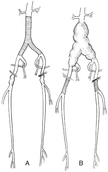
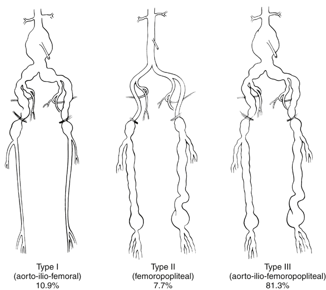
Arteriomegaly with aneurysms or aneurysmosis can be classified into three types based on the location and degree of arterial involvement. This classification, based on a review of 5771 aortofemoral angiograms performed at the Mayo Clinic from 1986 to 1982, revealed 300 patients with radiographic findings of arteriomegaly, including 91 (30.3%) with diffuse aneurysmal involvement or aneurysmosis (Figure 1).


Type I includes aneurysms of the aorta, iliac, and common femoral arteries with arteriomegaly of the superficial femoral and popliteal arteries. Type II includes aneurysms of the common femoral, superficial femoral, and popliteal arteries, with arteriomegaly of the aorta and iliac arteries. Type III includes aneurysms of the aorta, iliac, femoral, and popliteal arteries, with arteriomegaly of the intervening arteries that are not specifically aneurysmal.


Surgical Management


Surgical management is based on the clinical presentation of the disease and the extent of aneurysmal involvement. In the elective setting, all patients should have an angiographic study of the entire abdominal aorta with bilateral lower extremity runoff. It is also necessary to evaluate the aortic arch and thoracic aorta given the propensity for development of aneurysms in these locations. In the emergency setting, preoperative computed tomography angiography (CTA) with lower extremity runoff and intraoperative angiography can aid in planning the surgical reconstruction.


It is imperative to recognize that arteriomegaly and aneurysmosis represents a separate and distinct problem. The lack of any normal arterial segment can make it difficult to determine a suitable landing zone for both proximal and distal fixation of endovascular grafts. An exception exists in the case of type I aneurysmosis, where the external iliac artery is usually dilated but free of any aneurysmal changes. This affords the possibility of using an endograft with an appropriate amount of oversizing of the distal limbs. A similar opportunity exists for type II aneurysmosis, specifically with involvement of the distal femoral popliteal segment and in patients having generalized arteriomegaly and isolated popliteal artery aneurysms. With an appropriate amount of oversizing, it is feasible to place a covered stent graft across an aneurysmal popliteal artery in these cases. This course would be pursued only after thrombolysis of a thrombosed popliteal aneurysm and only in patients determined to be unsuitable or high-risk for open intervention.


Type I


The pattern of aneurysmosis in type I cases is similar to infrarenal aortic aneurysms with common iliac and femoral involvement. The external iliac artery is more often dilated but usually free of any aneurysmal changes, which permits either an open or endovascular intervention followed by femoral artery aneurysm resection with an expanded polytetrafluoroethylene (ePTFE) or Dacron interposition graft. In the patient with external iliac artery involvement, suitable endovascular options are limited and open repair is often indicated.


Open intervention for type I aneurysmosis is best treated in a single operation with aortic replacement in an end-to-end fashion with a bifurcated Dacron or ePTFE graft. The distal anastomosis is tailored to the degree of involvement of the external and hypogastric arteries. In patients with relatively normal external and hypogastric arteries, the distal anastomosis is performed by incorporating the origin of the external iliac artery and excluding the common iliac artery aneurysm.


Femoral involvement can be treated with an appropriate interposition graft. In cases with involvement of the external iliac and/or hypogastric artery, the distal anastomosis is performed to the bifurcation of the distal common femoral artery. Reimplantation of at least one hypogastric artery should be included with the repair, and proximal aneurysmal degeneration can be treated with a short interposition graft (Figures 2 and 3). The presence of a large or obviously patent inferior mesenteric artery, on which the sigmoid colon blood supply depends, is an indication that the inferior mesenteric artery might need to be reimplanted into the main body of the graft.


Stay updated, free articles. Join our Telegram channel

Aug 25, 2016 | Posted by in CARDIOLOGY | Comments Off on Arteriomegaly and Aneurysmosis

Full access? Get Clinical Tree

Get Clinical Tree app for offline access