Arteriographic Evaluation of Cerebrovascular Disease


Arteriographic Evaluation of Cerebrovascular Disease


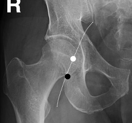
Thabele M. Leslie-Mazwi, Ronil V. Chandra, Daniel C. Oh, Albert J. Yoo and Joshua A. Hirsch


Catheter-based angiography is considered the gold standard for imaging the cerebral vasculature. The first report of x-ray angiography of vasculature was in 1896, performed in Vienna on a cadaveric specimen. The field has progressed dramatically since that time, with many important developments during the last 2 decades.


Indications for Catheter-Based Angiography


Diagnostic Indications


Catheter-based angiography provides a very high spatial resolution (200–300 μm) with excellent temporal resolution, making it invaluable in assessing primary neurovascular disease. Diagnostic indications include evaluation of intracranial hemorrhagic and ischemic disease, characterization of intracranial aneurysms, arteriovenous malformations (AVMs), dural arteriovenous fistulas, acute ischemic stroke, cerebral vasculopathy of various causes, and cerebral vasospasm. Diagnostic angiography in certain circumstances is followed by endovascular therapy.


Angiographic Procedures


Access to the Arterial System


The first step in catheter-based angiography of the cerebral vessels is safe arterial access. This is most typically accomplished through a transfemoral route. The common femoral artery is punctured at the femoral head (Figure 1).


The location of the puncture site is important because it allows effective compression of the arteriotomy site after the procedure. A puncture location too low (in the superficial femoral artery) risks inadequate postprocedural compression because of the absence of a firm structure to compress against, and a puncture too high (above the inguinal ligament) risks retroperitoneal hematoma formation. Transfemoral angiography is usually performed with a sheath but can be accomplished without one. By convention, the right groin is the site of access unless extenuating circumstances (e.g., previous right iliofemoral surgery, right iliofemoral atherosclerotic occlusion) are present, in which case the left groin is used.


The artery may be located by manual palpation for the femoral pulse and the underlying femoral head or, if location by palpation is difficult, with ultrasound guidance. Arterial puncture can be single or double walled and is typically accomplished using a micropuncture kit, and then the Seldinger technique is used to dilate the arteriotomy enough to allow the sheath to be inserted.


If femoral access is not an option because of the patient’s anatomy or comorbid disease, alternative routes of access may be used. The most common of these is a radial artery approach. This should be preceded by an Allen test to ensure adequate collateral circulation to the hand from the ulnar artery. Data suggest poor collateral circulation in the hand is present in approximately 23% of patients, and if it is noted with the Allen test, it is a reason to avoid the radial approach. The technique of access resembles the transfemoral approach, with placement of a 4 or 5 French (Fr) sheath. A spasmolytic medication (e.g., calcium-channel blocker) infused through the radial sheath is often required to prevent reactive spasm and possible thrombosis of the radial artery. A second alternative is a brachial artery approach, but because this artery is the sole vascular supply to the distal upper extremity, the consequence of access complications is more pronounced.


The advantages of the radial and brachial approaches include avoiding the risk of retroperitoneal hemorrhage and the need for bed rest associated with femoral artery puncture. In addition, an upper extremity approach may be useful for access to subclavian artery branches when vessel tortuosity prohibits a femoral approach. When an upper extremity approach is necessary, we favor the high brachial approach over the axillary approach because it provides a better opportunity for compression after the procedure.


Other, much less common options for arterial access include direct carotid artery puncture with sheath placement or a retroperitoneal lumbar stick, but these are rarely performed in current practice.


Sheath and Catheter Principles


The presence of a sheath allows rapid catheter exchange and decreases the risk of intraprocedural bleeding at the puncture site by minimizing trauma to the arteriotomy. Typically, a short sheath (10–13 cm) is used, though in the presence of significant iliofemoral tortuosity or atherosclerosis, sheath lengths up to 25 cm may be useful. Sheath diameters are measured where 1 Fr = 0.33 mm. The range of sheath sizes is from 4 Fr to 9 F for cerebral angiography, and the typical sheath size for diagnostic angiography is 5 Fr. It is important to note that the size refers to the inner diameter of the sheath and therefore determines what size catheter or device can be placed through the sheath. The outer diameter is approximately 1.5 to 2 Fr larger than the listed size and reflects the diameter of the actual arteriotomy.


Diagnostic catheters are inserted through the sheath. A wide range of catheters are available, with a variety of distal tip shapes to accommodate variations in vascular anatomy. Less distal catheter curvature is typically required in younger patients because of their more linear vasculature. In patients with markedly tortuous vessels, a reverse-curve catheter is required to access vessels off the aortic arch.


Diagnostic catheters are typically advanced over a hydrophilic guidewire. The guidewire allows atraumatic navigation of the catheter by keeping the catheter tip away from the wall of the vessel and therefore preventing dissection. A sufficient length of wire should protrude from the distal tip of the catheter to provide support for catheter advancement and to maximize the flexibility of the wire. If the distal wire protrudes only a short distance from the catheter tip, the exposed wire segment becomes exceedingly stiff and risks injury to the vessel. While catheters are measured in F, wire measurements are in thousandths of an inch (for example, a 0.038 wire is 0.038 inches in diameter).


Contrast Agents


A variety of different contrast agents are available for use in catheter-based angiography. Currently available iodinated contrast media can be divided into four classes with regard to their chemical structure: ionic monomers, ionic dimers, nonionic monomers, and nonionic dimers. The most important properties of these various agents are their viscosity, osmolality, solubility, hydrophilicity, and electric charge. The low-osmolal agents are less concentrated than conventional agents (600–850 mOsmol/kg versus 1500–1800 mOsmol/kg) but still are more concentrated than plasma. The iso-osmolal agents represent the more recent additions to the market. Low- and iso-osmolality contrast media are more comfortable for the patient and potentially cause less hemodynamic disruption than the conventional agents. However, these newer contrast media are significantly more expensive, which limits their widespread adoption.


Radiocontrast media can lead to acute kidney injury that begins soon after administration and is usually reversible. The nephrotoxic properties of these agents vary, and nonionic contrast agents are associated with a relatively decreased incidence of renal injury. The primary benefit of nonionic contrast agents in preventing renal dysfunction is seen in high-risk patients (e.g., serum creatinine ≥1.5 mg/dL or a glomerular filtration rate [GFR] <60 mL/min per 1.73 m2), particularly if the patient has diabetes (though the risk may still be as high as 25%). Therefore, these agents should be used instead of ionic high-osmolal agents in patients at risk. Additionally, contrast for hand injections can be diluted with saline to a 50:50 ratio to minimize its concentration.


Optimal therapy to prevent contrast-induced acute kidney failure remains uncertain but may include oral N-acetylcysteine (Mucomyst), usually dosed at 600 to 1200 mg twice daily for four dosages (two dosages before and two after the procedure), and intravenous sodium bicarbonate infusion, started 1 hour before and continued for 6 hours after the procedure. Patients with normal kidney function are at little risk, and few precautions are necessary other than avoiding volume depletion. In patients with near-normal kidney function, contrast doses up to 700 mL at iodine concentrations up to 300 mg/mL can be tolerated. For diagnostic cases that are anticipated to require many vessel injections (e.g., cerebrospinal angiogram), the total volume of contrast used should be monitored throughout the procedure to prevent excessive contrast administration.


Patients who have a confirmed or suspected allergic reaction to previous contrast exposure may still undergo cerebral angiography safely. However, they need to receive preprocedural medications. At our institution this involves prednisone 50 mg by mouth (PO) every 6 hours for three dosages ending 1 hour before the procedure and diphenhydramine 50 mg IV with ranitidine 150 mg IV 1 hour before the procedure, which helps to reduce the incidence of allergic reactions through mast cell stabilization and blockade of histamine receptors. In emergency patients where premedication is not possible, hydrocortisone 200 mg IV stat and repeated every 4 hours during the procedure is recommended. A thorough discussion of this specific risk should be initiated with the patient or the patient’s health care proxy before the procedure and informed consent should be obtained.


Vessel Selection


The cerebral angiogram should always be planned before the procedure. In general, it is easiest to begin with the right common carotid artery, then the left common carotid followed by the left vertebral artery (in left vertebral artery–dominant patients) to constitute a three-vessel cerebral arteriogram. Often, the intradural right vertebral artery can be visualized through the injection of the left vertebral artery, with the reverse being true in right vertebral artery–dominant patients. If the patient’s tolerance or cooperation is a concern, it is helpful to begin with the vessel of interest, because the procedure might need to be terminated early. All catheter navigation should be accomplished under direct fluoroscopic visualization. Discussion of individual vessel selection follows.


Right Common Carotid Artery


An angled-tip hydrophilic wire is advanced through the angled-tip diagnostic catheter that has been positioned in the descending thoracic aorta. The wire is advanced gently over the aortic arch, ensuring the tip of the wire is pointed inferiorly to avoid disrupting potential atherosclerotic plaque at the great vessel origins. The tip of the wire is positioned in the ascending aorta proximal to the expected origin of the brachiocephalic artery. The diagnostic catheter is advanced over the stationary hydrophilic wire, again ensuring that the tip of the catheter is angled inferiorly. Once the catheter nears the wire tip, the wire is brought back into the catheter, and the catheter is gently pulled back while rotating the tip to the right and superiorly to engage the brachiocephalic origin.


Once the diagnostic catheter is well seated in the vessel origin, the hydrophilic wire is advanced gently out of the catheter, with the tip oriented anteromedially to engage the right common carotid origin (the subclavian artery arises posterolaterally). The hydrophilic wire is advanced into the distal right common carotid artery, proximal to the carotid bifurcation (usually at the C4–5 disc level). It is helpful to look for calcified plaque at the carotid bifurcation to prevent the wire from crossing the bifurcation. The diagnostic catheter is then advanced over the wire.


The wire should be removed slowly, and a gentle hand puff of contrast should be used to confirm good catheter position and antegrade flow before injecting contrast for angiography.

Stay updated, free articles. Join our Telegram channel

Aug 25, 2016 | Posted by in CARDIOLOGY | Comments Off on Arteriographic Evaluation of Cerebrovascular Disease

Full access? Get Clinical Tree

Get Clinical Tree app for offline access