Arm Veins for Lower Extremity Arterial Reconstruction


Arm Veins for Lower Extremity Arterial Reconstruction


George Andros


Arm veins are now well established as versatile arterial conduits for infrainguinal arterial reconstruction. Although cephalic veins were demonstrated to be effective for leg revascularization in 1969, they did not receive wide application until the early 1980s. By that time it had become known that both the cephalic and basilic veins, either as single-length grafts or as part of an autogenous composite graft, had many advantages. They could be used if the saphenous vein was unavailable as a result of varicose vein stripping or if it was used for a previous cardiac or peripheral arterial bypass. They filled many roles: the need for multiple reoperations to achieve limb salvage, inflow and outflow extension grafts, and in sites where there was a risk of infection. The ability of autogenous grafts to tolerate low flow made them attractive for bypass to infrapopliteal and paramalleolar arteries. Despite the burgeoning of endovascular techniques for leg revascularization, autogenous conduits continue to occupy an important place in complex leg revascularization procedures.


Anatomy


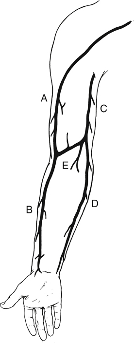
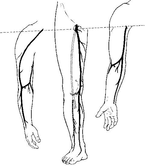
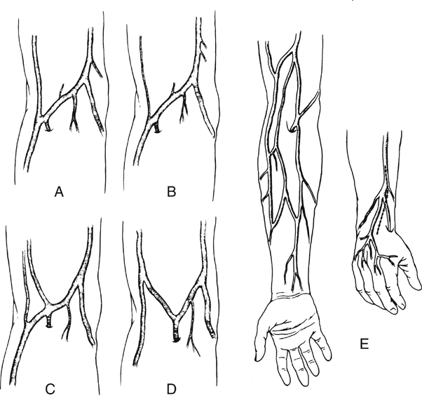
The cephalic vein arises at the anatomic snuff box of the wrist, courses on the lateral aspect of the arm and upper arm, continues in the deltopectoral groove, and ends in its confluence with the axillary vein. It is nearly uniform in diameter (5–7 mm) and is best used in the reversed configuration (Figure 1).


The medially positioned basilic vein originates near the ulnar styloid process, passes anterior to the median condyle of the elbow, and widens as it terminates in the brachial or ultimately the axillary vein. At its largest end it measures 7 to 12 mm in diameter, tapering down to 3 to 5 mm (see Figure 1). The upper arm basilic vein normally joins one of the brachial veins near the axilla, although this juncture can occur anywhere along its course in the upper arm. One of the brachial veins can substitute for the basilic vein by arising as a direct continuation of the median antecubital vein. The median antecubital vein (MAV) interconnects the cephalic and basilic veins, is devoid of valves, and has numerous large, deep branches (see Figure 1). The entire basilic vein can span from the common femoral artery to the popliteal artery, whereas the cephalic vein is long enough to reach crural arteries in the midcalf (Figure 2).


Two naturally occurring continuous grafts are also available in addition to the cephalic and basilic veins. The first consists of the forearm cephalic, median antecubital, and upper arm basilic segments (see Figure 1). We have used this graft reversed as well as nonreversed. When reversed, this graft usually tapers less than a reversed saphenous vein graft. A second continuous graft takes advantage of the segments with the largest diameters: the upper arm basilic, median antecubital, and upper arm cephalic veins (see Figure 1). The basilic vein is oriented at the proximal anastomosis because its diameter is opposite to the proximal artery. However, we have used this single-length segment successfully in less than half of the intended attempts. This low rate of use is the result of intimal damage and fibrous synechia within the median antecubital vein that often accompanies fibrosis of the adjacent forearm cephalic vein. Resection of the MAV and end-to-end anastomosis of the upper arm cephalic and basilic segments yields a graft that is 4 to 5 inches shorter. If a composite graft must be surgically constructed, the cephalic vein segment should be placed distally because of its smaller diameter.


There are many anatomic variants of the arm veins, principally in the antecubital fossa and the forearm. The normal anatomy can vary, with one to four or more deep muscular branches occurring in the region of the cephalic–median antecubital juncture (Figure 3). A small upper arm cephalic vein is usually associated with an enlarged median antecubital vein connecting to the basilic vein (see Figure 3). Skin incisions must be placed precisely if there is duplication of the upper arm cephalic and forearm or wrist cephalic veins (see Figure 3). Convergence of the cephalic and basilic veins in the antecubital fossa to form a Y was thought to preclude the use of the aforementioned single-length upper arm loop grafts. This has not been a problem because the angles become unkinked under arterial pressure.


Preoperative Assessment


Visual inspection is the first step in the preoperative evaluation of arm veins. Tourniquets, dependency, hot packs, and exercise in a warm room are helpful adjuncts as the arms are examined for evidence of palpable and compliant veins free of thrombosis, fibrosis, and discontinuity. The skin is inspected for signs of recent or repeated puncture. If visual inspection fails to demonstrate a satisfactory vein, duplex ultrasonography is the preferred technique for preoperative arm vein assessment. Ultrasonography is effective and noninvasive, and it eliminates the risk of contrast-induced phlebitis that is associated with phlebography.


Assessment of arm veins as possible bypass grafts is indicated for patients with varicose veins, a history or clinical evidence of deep or superficial venous thrombosis, past vein stripping, vein harvesting, or the possibility of vein damage caused by previous operations in the lower extremity and elsewhere. Multiple previous illnesses, operations, and hospitalizations often leave patients with vein damage induced by intravenous infusions. Such a history is likewise an indication for arm vein imaging. We are inclined not to subject the patient to vein mapping if the ipsilateral greater saphenous vein is undisturbed or arm veins appear to be acceptable by visual inspection. Duplex imaging of arm veins should be selective rather than routine.


Color Doppler imaging includes assessment of diameter and continuity of the lumen using high-frequency probes while incorporating an offset or a wedge if the vein is immediately under the skin. The ultrasonographically determined internal diameter of the in-situ arm vein underestimates the outside diameter of the arterialized vein, on average by 2 mm. Veins with diameters of less than 2 mm, discontinuities, or hyperechoic areas are usually not usable. Noncollapsibility of veins under probe compression suggests thrombosis. Vein compressibility, however, does not rule out mural damage.


One should not overlook the arm veins in patients whose arms are so obese that the veins cannot be judged by physical examination. The increased prevalence of obesity in the United States and other developed countries has made visual inspection less dependable for preoperative assessment and has led to greater reliance on ultrasonography The fat that obscures the vein also shields it from venipuncture.


No differences exist between the arm veins of men and women as regards relative length, diameter, strength, or clinical use. The creation of a cephaloradial arteriovenous fistula to arterialize the vein before its translocation as a bypass graft has been described. We have not used this technique and doubt that it is necessary or even feasible in most cases. Perioperative angioscopy of the arm veins has been shown to be an important adjunct to improve the usability of arm veins. The condition of the vein and the presence of mural defects can be evaluated in some instances without the need to unroof or harvest the vein. Long fibrotic and scarred segments must be resected, but minor synechiae occasionally can be managed through the angioscope. Although it is not always possible, most experienced users of arm veins, as with leg veins, advise that the condition of the vein and its usability be determined during the unroofing process before the vein is harvested.


Indications and Contraindications for Arm Vein Grafts


Although options for leg revascularization are now abundant, the proven current treatment for long occlusions remains a bypass graft. The list of bypass conduits, headed by autogenous materials, includes ipsilateral greater saphenous vein, in situ, reversed, or nonreversed; contralateral greater saphenous vein, reversed or nonreversed; arm vein; short saphenous vein; and synthetic materials such as knitted Dacron or expanded polytetrafluoroethylene (ePTFE) conduits (with or without distal vein cuffs).


Of the arm veins, the order of preference for the author’s group is cephalic vein; forearm cephalic–median antecubital–basilic vein; nonreversed valve incised basilic vein; basilic–median antecubital–upper arm cephalic vein; and composites of autogenous venous segments.


If the ipsilateral saphenous vein is absent or inadequate and if there is evidence of contralateral leg ischemia, we are inclined to use arm veins. Prior deep vein thrombosis can require preservation of the greater saphenous vein as an important venous effluent channel. Postoperative leg edema is reduced by using arm veins, short incisions, and a superficial graft tunnel.


Gangrene, nonhealing ulcers, rest pain, and severe claudication are all appropriately managed with arm vein bypass grafting. The use of arm veins is not restricted to limb-threatening ischemia. The infrageniculate popliteal, crural, perimalleolar, and plantar arteries are suitable distal anastomotic sites. Long occlusions of the upper extremity arteries are readily bypassed with arm veins; the ipsilateral cephalic vein from the arm receiving the bypass is selected if saphenous vein is unavailable. Arm veins, preferably the basilic vein because it is somewhat more robust, effectively substitute for saphenous veins as a bypass between the carotid and axillary arteries. Renal artery bypasses have been performed with basilic veins, but rather than using the aorta, we use the celiac or splenic artery for the proximal anastomosis. By analogy, cardiac surgeons at our institution have chosen to originate coronary artery arm vein bypasses from vessels other than the aorta.


Despite this long list of indications for the use of arm veins, patching of arterial defects is not among them. There is uncertainty whether an arm vein patch can withstand arterial pressure without becoming aneurysmal, and, hence, this application has been avoided.


Venous disease is an emerging indication for vascular reconstruction. Successful operative management of the post-thrombotic syndrome by valve transplantation depends on the availability of a large-diameter, valve-bearing segment of axillary vein. We have not substituted axillary veins for a spiral saphenous vein graft in the management of superior vena caval syndrome but believe that it may be a promising application. In contrast to arterial patches, basilic veins make ideal patches for venous defects, and they have been used to reconstruct the renal vein, vena cava, and femoral vein.


Protecting and preserving the arm veins are indispensable measures to ensure operative success. The patient, nurses, other paramedical personnel, and physicians, especially anesthesiologists, are admonished against venipuncture and insertion of intravenous catheters above the wrist. The veins are protected preoperatively by placing the arms in gauze wraps, on which is written, “No Venipunctures or IVs.” Warnings are placed prominently in the chart and in the patient’s room. Nursing and paramedical personnel participate in an educational program on the importance and method of preserving arm veins.

Stay updated, free articles. Join our Telegram channel

Aug 25, 2016 | Posted by in CARDIOLOGY | Comments Off on Arm Veins for Lower Extremity Arterial Reconstruction

Full access? Get Clinical Tree

Get Clinical Tree app for offline access