Approach to the Diagnosis of Nonneoplastic Parenchymal Lung Nodules


Approach to the Diagnosis of Nonneoplastic Parenchymal Lung Nodules


Allen P. Burke, M.D.

Marie-Christine Aubry, M.D.

Haresh Mani, M.D.


Poorly Formed Granulomas

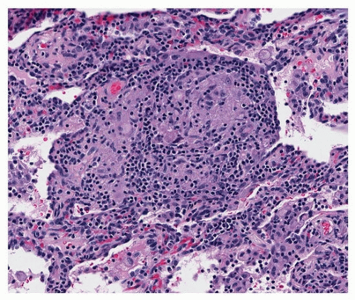
Granulomas in the lung parenchyma are generally classified as poorly or well formed. Poorly formed granulomas are a hallmark of hypersensitivity to inhaled allergens. They are indistinct collections of lymphocytes with macrophages, typically giant cells. Poorly formed granulomas are generally near the bronchovascular bundles and are often associated with interstitial inflammation (Fig. 9.1).


FIGURE 9.1 ▲ Poorly formed granuloma. The interstitium contains chronic inflammation with granulomatous inflammation in the center (epithelioid macrophages with a few giant cells).


Well-Formed Granulomas

Well-formed granulomas are found in sarcoid or infections, usually fungal or mycobacterial. sarcoid and infections (Table 9.1). There is a compact collection of epithelioid histiocytes with a discrete border (Fig. 9.2) and a subpopulation of T lymphocytes. Sarcoid granulomas are found in a lymphatic distribution in the bronchovascular bundles, interlobular septa, and subpleural space. Fibrosis is a characteristic of sarcoid granulomas (Fig. 9.3). Infectious granulomas are, in contrast, asymmetric and occur randomly in the parenchyma. Further, presence of significant lymphoplasmacytic inflammation favors an infectious etiology. Well-formed granulomas of any cause, but especially sarcoid, may contain mineral deposits (Schaumann bodies), which should not
be mistaken for pneumoconiosis (Fig. 9.3). Asteroid bodies are also nonspecific features of well-formed granulomas (Fig. 9.4).


TABLE 9.1 Histologic Features of Pulmonary Granulomas


Diagnosis


Necrosis


Multiplicity


Contour


Lymphatic Distribution


Neutrophils


Multinucleated Macrophages


Necrotizing Vasculitis


Distinguishing Features


Sarcoidosis


10%-20%a


100%


Regular (100%)


70%-100%


Absent


100%


Absent


Diffuse, symmetric


Mycobacterial infectionb


75%


90%


Regular (2/3)


20%


80%


100%


Absent


Positive AFB stain (about 50%)


Histoplasmosis


>90%


75%


Regular (80%)


<5%


50%


80%-90%


Absent


Positive GMS stain (>90%, but requires careful review)


Coccidioidomycosis


>90%


80%


Regular (90%)


<5%


90% also, frequent eosinophils


90%


Absent


Positive GMS stain (>90%)


Blastomycosis


>90%


80%


Irregular (75%)


<5%


100%


50%-80%


Absent


Positive GMS stain (>90%)


Talc granulomas


Absent


100%


Regular


Absent


Infrequent


100%


Absent


Birefringent crystals


Granulomatosis with polyangiitis


100%


>90%


Irregular (100%)


0


100% (usually with apoptosis)


60%


20%-50%


Apoptotic neutrophils causing basophilic necrosis


Idiopathic (unexplained)


>95%; abundant in 75%c


80%


Regular (60%)


<20%


50%


80%


Absent


Are presumed infections; some will demonstrate NTM DNA by PCR


Rheumatoid nodule


100%


60%


Irregular (60%)


0


80%


100%


Absent


History and serology


Hyalinizing granuloma


Absent


50%


Regular


Absent


Absent


Absent


Absent


IgG4-related histologic features


Percentages are approximate. For further histologic features distinguishing fungal infections, see Chapter 41 and 44.


a Necrosis in sarcoid is usually inconspicuous and central in the granuloma.


b Nontuberculous (atypical) mycobacterial granulomas are more frequently encountered as lung nodules than tuberculosis. Histologic features are different in infections in immunocompromised patients, and discrete nodules are not as common (see Chapter 40).


c Idiopathic nonnecrotizing granulomatous disease is synonymous with sarcoidosis and is typically bilateral and symmetric. Solitary idiopathic necrotizing granulomas are common. Well-formed granulomas that do not have necrosis and that form solitary lung nodules are rare.


NTM, nontuberculous mycobacteria; PCR, polymerase chain reaction.


Adapted in part from Mukhopadhyay S, Wilcox BE, Myers JL, et al. Pulmonary necrotizing granulomas of unknown cause: clinical and pathologic analysis of 131 patients with completely resected nodules. Chest. 2013;144:813-824.

Only gold members can continue reading. Log In or Register to continue

Stay updated, free articles. Join our Telegram channel

Aug 19, 2016 | Posted by in CARDIOLOGY | Comments Off on Approach to the Diagnosis of Nonneoplastic Parenchymal Lung Nodules

Full access? Get Clinical Tree

Get Clinical Tree app for offline access