Aortorenal Bypass for Renovascular Hypertension in Adults


Aortorenal Bypass for Renovascular Hypertension in Adults


Kimberley J. Hansen, MD


The introduction of new, highly potent antihypertensive agents and percutaneous intervention has changed many attitudes regarding open surgical treatment for renovascular disease. Many physicians recommend surgical intervention for severe hypertension despite maximal medical therapy, for failures or disease patterns not amenable to percutaneous dilation, or for renovascular disease associated with excretory renal insufficiency (ischemic nephropathy).


Preoperative Preparation


Antihypertensive medications are reduced during the preoperative period to the minimum necessary for blood pressure control. Many patients who require large doses of multiple medications for control have reduced requirements while hospitalized and on bed rest. If continued antihypertensive therapy is required, peripheral vasodilators (e.g., amlodipine) and selective β-adrenergic blocking agents (e.g., atenolol or metoprolol) are the agents of choice. There are few adverse hemodynamic effects when these agents are combined with general anesthesia. If an adult’s diastolic blood pressure exceeds 120 mm Hg, it is essential that operative treatment be postponed until the blood pressure is controlled. In this instance, the combination of intravenous nitroprusside and esmolol is administered in an intensive care setting with continuous intraarterial blood pressure monitoring. Similarly, if the patient has significant heart disease, pulmonary artery wedge pressure, cardiac index, and oxygen delivery are monitored to optimize cardiac performance before and after operation. Information about regional myocardial performance and left ventricular preload is provided by transesophageal echocardiography in high-risk cardiac patients.


Although new diagnostic imaging techniques might have future application, digital subtraction aortography remains an essential diagnostic study before renal artery reconstruction. This is particularly important in planning branch renal artery bypass. Arteriography better defines the distal extent of renal artery disease and intrarenal disease consistent with nephrosclerosis. Although 70% of the patients treated at the author’s center have at least mild ischemic nephropathy, no patient has been made permanently dependent on dialysis after digital subtraction arteriography.


Finally, certain measures are used in almost all renal artery operations. Mannitol is administered intravenously in 12.5-g doses early in the operation. Repeated doses are administered before and after periods of renal ischemia, up to a total dose of 1 g/kg body weight. Just before renal artery cross clamping, unfractionated heparin sodium, 100 U/kg, is given intravenously, and systemic anticoagulation is verified by measuring activated clotting time. Unless required for hemostasis, protamine is not routinely administered to reverse the heparin effect at the completion of the operation.


Operative Management


Surgical Strategy


A surgical strategy has evolved for open renal artery reconstruction (Box 1). Hypertension is a prerequisite for renal artery intervention by any method. Management of most unilateral lesions is guided by results of functional studies. Empirical renal artery repair is often performed without functional studies in patients with severe hypertension, especially when hypertension is associated with renal insufficiency and bilateral renal artery lesions. Prophylactic renal artery repair in the absence of hypertension is not recommended, either as an isolated procedure or combined with aortic reconstruction.


During surgical intervention, all hemodynamically significant renovascular disease is corrected in a single operation. However, if bilateral ex vivo reconstruction is required, a staged approach is planned. Although kidney size, evidence of preoperative function, and renal histology have been suggested as measures to guide renal artery reconstruction as opposed to nephrectomy, the author has reserved nephrectomy for unreconstructible renal artery disease to a nonfunctioning kidney. When the renal artery distal to an occlusive lesion is grossly normal, the author favors revascularization over nephrectomy, having observed beneficial blood pressure and renal function response regardless of kidney size or histologic pattern on renal biopsy.


The majority of operative renal artery repairs (98%) at the author’s center use direct aortorenal methods of reconstruction. In part, this reflects the current demography, in which 40% of patients have concomitant visceral artery disease. Consequently, only 1% of renal artery reconstructions use indirect methods or splanchnorenal bypass. Regardless of the method of repair, each reconstruction is studied during operation with renal duplex sonography. Major B-scan defects are revised before the procedure is completed, reducing the rate of early postoperative failure to less than 0.5%.


Operative Exposure


A xiphoid-to-pubis midline abdominal incision is made for operative repair of atherosclerotic disease of the main renal artery, especially when bilateral lesions are present. The last 1- or 2-cm cephalad incision coursing to one side of the xiphoid is important in obtaining full exposure of the upper abdominal aorta and renal artery origins. Some form of fixed mechanical retraction is also advantageous, particularly when combined aortorenal procedures are required. Otherwise, extended flank and subcostal incisions are reserved for fibrodysplastic lesions, and splanchnorenal bypass and combined mesenteric reconstruction and branch reconstruction require ex vivo techniques.


When left renal bypass is combined with antegrade mesenteric bypass, an extended flank incision is useful. With the left flank elevated, the incision extends from the right semilunar line into the flank, bisecting the abdominal wall between the left costal margin and iliac crest. A left visceral mobilization allows access to the renal and mesenteric vasculature. If necessary, the aortic crus can be divided, and an extrapleural dissection of the descending thoracic aorta can provide access to the T9 to T10 thoracic aorta for proximal control.


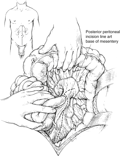
When the midline xiphoid-to-pubis incision is used, the posterior peritoneum overlying the aorta is incised longitudinally, and the duodenum is mobilized at the ligament of Treitz (Figure 1). During this maneuver, it is important to identify mesenteric arterial collaterals that course at this level. Finally, the duodenum is reflected to the patient’s right to expose the left renal artery. By extending the posterior peritoneal incision to the left along the inferior border of the pancreas, an avascular plane posterior to the pancreas can be entered (see Figure 1) to expose the entire renal hilum.


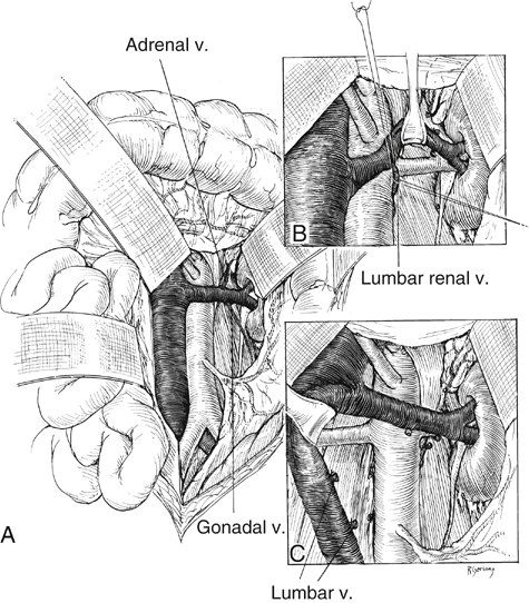
This exposure is of special significance when distal renal artery lesions need to be managed (Figure 2A). The left renal artery lies posterior to the left renal vein. In some instances, the vein can be retracted cephalad to expose the artery; in others, caudal retraction of the vein provides better access. Usually, the gonadal and adrenal veins, which enter the left renal vein, must be ligated and divided to facilitate exposure of the distal artery. Often, a lumbar vein enters the posterior wall of the left renal vein and can be easily avulsed unless special care is taken while mobilizing the renal vein (Figure 2B). The proximal portion of the right renal artery is exposed through the base of the mesentery by ligating two or more pairs of lumbar veins and retracting the left renal vein cephalad and the vena cava to the patient’s right (Figure 2C).


Stay updated, free articles. Join our Telegram channel

Aug 25, 2016 | Posted by in CARDIOLOGY | Comments Off on Aortorenal Bypass for Renovascular Hypertension in Adults

Full access? Get Clinical Tree

Get Clinical Tree app for offline access