Aortoiliac Occlusive Disease
HUITING CHEN and JONATHAN L. ELIASON
Presentation
A 53-year-old female presents to the clinic for worsening lifestyle-limiting back and thigh pain with ambulation for several years, relieved by rest, and nonhealing ulcers to her lower extremities. Her past medical history includes diabetes mellitus, deep venous thrombosis and pulmonary embolus, hyperlipidemia, hypertension, and pyoderma gangrenosum, with significant wound-healing difficulties. Her medications include daily aspirin 81 mg, a low-dose beta-blocker, Coumadin, and a statin. Her past surgical history includes an angioplasty of her superficial femoral arteries and an inferior vena cava filter. She has a current tobacco use history of two packs per day for 30 years. On physical examination, her blood pressure is 135/58 and her pulse 84. Bilateral femoral and distal pulses are nonpalpable, though distal signals are present by Doppler. Her lower back, buttock, and thighs exhibit multiple shallow coin-sized superficial ulcerations in a bilateral distribution.
Differential Diagnosis
Causes of lower extremity pain with exertion may be arterial, venous, neurogenic, arthritic, or due to diabetic neuropathy. The presentation of symptoms is different for these varied etiologies, and often, the history will allow sufficient narrowing of the differential diagnoses. Arterial claudication is due to demand ischemia with exertion and is classically described as cramping or aching with walking a certain distance, consistently relieved with rest. Venous claudication is due to venous occlusion, with subsequent “bursting” sensation in the legs and associated lower extremity edema. Neurogenic causes of leg pain include sciatic pain from lumbosacral nerve root compression, often presenting with diffuse pain extending from the buttocks to the feet. This pain is classically improved with bending at the waist, which relieves the nerve compression from spinal stenosis. Arthritic pain is experienced at joint spaces and not classically associated with onset of a consistent walking distance. It may be improved with activity and worse just after getting up. Lastly, diabetic neuropathy is often associated with structural foot changes and poorly healing ulcers in the setting of a history of diabetes.
In patients with peripheral arterial disease (PAD) due to inflow insufficiency, the level of disease is found at the aorta or iliac arteries. Patients with aortoiliac occlusive disease (AIOD) will often have diminished or absent femoral pulses and distal pulses possibly only detectable by Doppler.
Diagnostic Tests and Result
Ankle-Brachial Index
The ankle-brachial index (ABI) is easily obtained in a clinical setting by the use of a blood pressure cuff and a Doppler probe. The cuff is affixed to the subject’s assessed leg, as close to the ankle as possible. The probe is placed over the location of the dorsalis pedis artery on the dorsum of the foot or over the posterior tibial artery posterior to the medial malleolus while the cuff is inflated above systolic pressure. While the cuff is slowly deflated, the blood pressure for the dorsalis pedis and posterior tibial arteries is measured and recorded when return of arterial Doppler signals is obtained. The higher of these two distal systolic pressures is then divided by the systolic pressure obtained from the brachial artery to obtain the ABI. Normal ABIs range from 0.9 to 1.29, with decreases in value as the severity of PAD increases. Mild to moderate PAD produces ABIs of 0.4 to 0.9. Rest pain, and possibly tissue loss, is typically associated with ABIs less than 0.4. Notably, calcification of the tibial vessels is often seen in diabetic patients, resulting in noncompressible vessels and falsely elevated ABIs. In these patients, often the toe-brachial index (TBI) more reliably reflects the extent of disease, as smaller digital vessels are less affected by the calcification process in diabetes.
Doppler Waveforms
Waveform analysis follows the principle that the waveform produced in a vessel is altered by stenosis in the vessel. Using a Doppler probe, normal waveforms have a triphasic character, with a brisk upstroke in systole, followed by a brief retrograde early diastolic peak, then a smaller antegrade diastolic peak. Mild stenosis produces a biphasic, widened waveform, due to loss of vessel recoil in diastolic flow. Peak systolic velocities also increase with increasing severity of stenosis. Obtaining Doppler waveforms from successive levels in the lower extremity is a useful adjunct in defining the anatomic level of disease. A monophasic waveform at the groin or femoral level in the right clinical context will strongly suggest the presence of AIOD.
Case Continued
The patient’s ABIs were 0.38 on the right and 0.39 on the left. Waveform analysis revealed monophasic morphology in the bilateral femoral arteries and biphasic flow in the tibial and pedal arteries. The combination of findings from physical examination, ABIs, and Doppler waveforms suggests AIOD. In patients whose symptoms are only mild to moderate claudication, conservative management with pharmacology, tobacco cessation, and a supervised exercise program may be attempted first. However, this patient’s symptoms are lifestyle limiting, and more extensive diagnostic studies with possible intervention are warranted. A CT angiogram (CTA) is preferred to catheter-based angiography in this nonacute setting, as the CT images the entire aortoiliac system without the risks associated with invasive testing.
CTA Abdomen/Pelvis: CTA from the referring hospital (images not shown) revealed total occlusion of the distal abdominal aorta and common iliac arteries bilaterally extending from just below the inferior mesenteric artery to the common iliac artery bifurcation on both sides. There is reconstitution of both internal and external iliac arteries from collateral vessels.
Diagnosis and Treatment
This patient has clinical symptoms of AIOD confirmed by noninvasive studies including a CTA demonstrating complete occlusion of the aorta and common iliac arteries bilaterally. In addition to a discussion of the risks and benefits of surgery, the critical importance of tobacco cessation is emphasized. The patient agrees to quit smoking and elects to proceed with an intervention.
Approach: Surgical versus Endovascular
The Trans-Atlantic Inter-Society Consensus (TASC) classification of AIOD by lesion morphology recommends endovascular therapy for the treatment of localized disease (TASC A and B), whereas extensive diseases (TASC C and D) are best approached with open reconstruction. Our patient with infrarenal aortoiliac occlusion has a TASC D lesion, which is typically best suited for surgery. The most commonly used and safe surgical approach to this type of TASC D lesion is aortobifemoral bypass. This operation may be performed in an end-to-side or end-to-end fashion for the proximal anastomosis onto the aorta, and distal anastomoses to the femoral arteries are performed in an end-to-side fashion. The distal anastomoses are constructed based upon the pattern of disease and anatomic lie of the graft limbs, with “hooding” the graft limb from the common femoral artery onto the profunda femoris artery being a common and effective reconstructive technique. The durability for this type of bypass procedure is high, with studies commonly reporting greater than 85% primary patency rates at 5 years. It is effective in relieving symptoms due to aortoiliac obstruction. It has long been considered the gold standard for treatment of severe aortoiliac disease.
However, recent advancements in devices and techniques have resulted in admirable outcomes for endovascular approaches to extensive disease. Taurino and colleagues reported that in 50 patients with TASC C or D lesions treated with either a hybrid approach or endovascular only, 100% achieved technical success, with no significant difference in cumulative primary or secondary patency rates between the two approaches. While not stratified by TASC levels, in comparing 118 aortofemoral bypass (AFB) patients with 174 aortoiliac angioplasty and stenting (AS) patients, Burke et al. found no difference in mortality, cerebrovascular accidents, myocardial infarction, or renal failure requiring hemodialysis. More recently in 2013, Sixt et al. evaluated 1184 patients of all four TASC classifications and noted no difference in restenosis, reintervention, or primary and secondary patency rates in the four subgroups at 12 months. While they found that the TASC D group required acute reintervention more often (p < 0.001), they concluded that for AIOD, the endovascular approach can be considered for treatment regardless of TASC classification.
In lesions with aortic occlusions, the addition of transbrachial access to standard femoral access has been reported in the literature. Bjorses et al. treated 173 patients of TASC A through D aortoiliac lesions, 11 of whom were recanalized due to aortic occlusions. Similar to our approach, these authors performed bilateral femoral and brachial access for these lesions and reported no statistically significant difference in primary patency between the TASC classes. Lagana and colleagues report endovascular treatment of 19 patients with Leriche syndrome, 5 of which have complete occlusion of the infrarenal aorta and both common iliac arteries. All of these five patients underwent brachial and femoral access; however, crossing of the lesion was not successful in one of these patients, who subsequently underwent an axillobifemoral bypass. Similar access for chronic aortoiliac occlusion by Moise resulted in successful endovascular reconstruction of the occlusion in 29/31 patients. Like Lagana’s approach, Moise’s patient for which the occluded aorta could not be traversed underwent an axillobifemoral bypass.
While both open and endovascular approaches were offered as treatment options, due to the patient’s diagnosis of pyoderma and history of poor wound healing, an endovascular approach was selected.
Endovascular Management
The bilateral common femoral arteries and left brachial arteries were percutaneously accessed under fluoroscopic and ultrasound guidance with a micropuncture needle. Using Seldinger technique, 6-French 25-cm-long Pinnacle sheaths were placed into the common femoral arteries, and a 5-French short Pinnacle sheath was placed in the left brachial artery. The patient was then systemically heparinized.
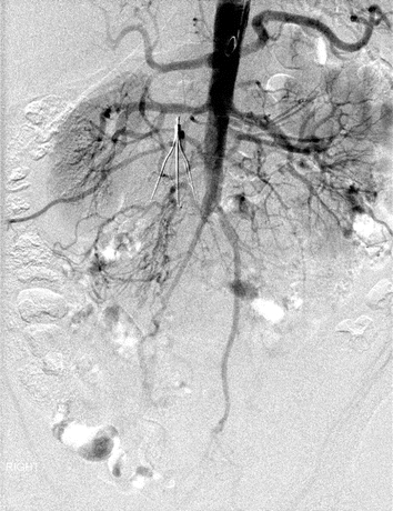
An aortogram was obtained from antegrade catheter advancement into the abdominal aorta, revealing large paired lumbar arteries providing collateral circulation to the pelvis. The aorta was occluded at the level of the patent origin of the inferior mesenteric artery with a completely occluded terminal aortic segment and bilateral common iliac arteries (Fig. 1). Reconstitution of the iliac arteries occurred at the iliac bifurcation. The length of occlusion was estimated to be between 90 and 100 mm.

FIGURE 1 Antegrade transbrachial abdominal aortogram revealing aortic occlusion at the level of the inferior mesenteric artery.


