Aortofemoral Bypass for Atherosclerotic Aortoiliac Occlusive Disease


Aortofemoral Bypass for Atherosclerotic Aortoiliac Occlusive Disease


David C. Brewster


Aortoiliac disease (AOID) is usually segmental in distribution and therefore amenable to effective treatment. Even in patients with multilevel disease, successful correction of hemodynamically significant inflow disease often provides adequate revascularization of the extremities and satisfactory clinical relief of ischemic symptoms.


Procedure Selection


Numerous options exist for revascularization in patients with AOID. Selection of the most appropriate method depends largely on two factors: the patient’s surgical risk and the extent and distribution of occlusive disease. Aortobifemoral grafting is elected for patients who are relatively free of serious comorbid medical conditions that would make them a high or prohibitive risk for direct abdominal aortic reconstruction. Thus careful preoperative evaluation is important.


For patients with relatively limited areas of disease, particularly unilateral iliac disease, alternative lesser procedures such as percutaneous transluminal angioplasty (PTA) with or without stenting, femorofemoral bypass, or unilateral iliofemoral grafting may be considered. For high-risk patients with bilateral iliac disease or those with relative contraindications to direct aortic reconstruction as a result of technical considerations, such as heavy retroperitoneal scarring or contamination, axillobifemoral extra-anatomic bypass may be chosen. The lower long-term patency rate of these grafts is accepted as a compromise to achieve revascularization in high-risk situations.


Although all of the alternative methods may be helpful or appropriate choices in selected circumstances, aortobifemoral grafting clearly provides superior long-term results in terms of durability and sustained relief of symptoms and thus should be properly regarded as the preferred treatment or gold standard for the management of atherosclerotic AOID, as well as the yardstick to which the results of alternative therapies must be measured.


Aortoiliac endarterectomy formerly was employed in many such patients. Although excellent results may be obtained in patients whose AOID is confined to the distal aorta and common iliac vessels, the majority of patients have diffuse disease and are better managed with aortofemoral graft insertion, which is more expedient and effective in such circumstances.


Operative Management


A broad-spectrum prophylactic antibiotic, such as cefazolin 1 g, is administered intravenously 1 to 2 hours before the operation and continued for 1 or 2 days postoperatively. In patients with an infected open ischemic lesion of an extremity or any other possible source of bacteremia, culture-specific antibiotics are best started several days before the operation.


A radial artery cannula is inserted in all patients for continuous blood pressure monitoring and arterial blood gas determinations. A Swan–Ganz catheter is used in many, but not all, patients, depending on preoperative assessment of cardiac and renal status. Most patients undergoing aortic reconstruction on the author’s vascular unit and in many other centers undergo anesthesia by a combination of epidural narcotics and inhalation agents (combined general and epidural technique). Continuation of epidural analgesia in the early postoperative period for pain control has been a significant advance in limiting administration of systemic narcotics and in reducing associated respiratory complications following aortic reconstruction.


Operative Approach


The infrarenal abdominal aorta may be exposed for aortofemoral reconstruction by a variety of approaches. A long midline vertical incision is employed most often and is generally preferred because it is rapidly made, is easy to close, and affords maximal exposure and technical flexibility in most patients. A retroperitoneal approach may also be used, if desired. For this method, the patient is positioned with the shoulders and torso rotated approximately 45 degrees toward the right, while the hips and extremities are maintained as horizontal as possible to facilitate exposure of the femoral arteries in each groin. An oblique left flank incision beginning at the tip of the twelfth rib and carried toward the midline just below the umbilicus is used.


A retroperitoneal approach is potentially advantageous in patients who have had multiple prior abdominal operations or, particularly, for reoperative aortic procedures. This approach is also often useful for obese patients or those with right-sided intestinal stomas. Advocates also believe that a retroperitoneal approach causes less postoperative ileus, decreased fluid losses, and reduced cardiopulmonary stresses postoperatively. However, many of these potential benefits are not conclusively established. The author prefers a transperitoneal midline approach, primarily because of the easier and generally superior exposure of the femoral arteries that it allows. A retroperitoneal approach is reserved for some of the specific technical indications outlined earlier.


Bilateral groin incisions are made to expose both femoral arteries. Any lymph nodes or lymphatic tissue is best divided between clamps and then suture ligated to minimize the possibility of a postoperative lymphatic leak with its associated risk of wound or graft infection. The posterior aspect of the inguinal ligament is partially divided directly over the femoral artery to ensure ample space for tunneling the graft without compression. Dissection is carried to just beyond the femoral artery bifurcations, and the proximal aspect of both superficial and deep femoral artery branches are encircled with silicone elastic (Silastic) loops. If preoperative arteriograms or palpation at the time of operation suggests significant occlusive disease involving the proximal deep femoral artery branch, more distal exposure of this vessel for at least another 2 to 3 cm is required to allow concomitant profundaplasty at the time of distal graft anastomosis. This method usually necessitates dividing and ligating one or more branches of the deep femoral vein that typically cross the anterior surface of the proximal profunda femoris artery.


Tunnel Construction


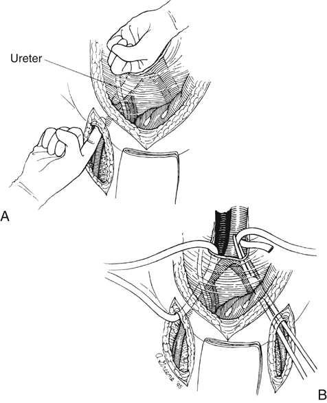
After the aortic and femoral artery dissection are completed, retroperitoneal tunnels between the two fields of dissection are made for subsequent passage of each graft limb. Such tunnels are best made by gentle blunt dissection with the index finger of each hand simultaneously from the groins and the area of the aortic bifurcation (Figure 1). Dissection should be kept on a plane directly anterior to the common and external iliac vessels to ensure that the graft is subsequently placed posterior to the ureter. This detail is important because passage of graft anterior to the ureter can lead to later compression and obstruction of the ureter and hydronephrosis. After appropriate tunnels have been created to both groins, a long blunt-tipped clamp is placed through the tunnel, and a Penrose drain is drawn through the tunnel. Elevation on both ends of this facilitates later passage of the graft limbs to the site of distal graft anastomosis.


Stay updated, free articles. Join our Telegram channel

Aug 25, 2016 | Posted by in CARDIOLOGY | Comments Off on Aortofemoral Bypass for Atherosclerotic Aortoiliac Occlusive Disease

Full access? Get Clinical Tree

Get Clinical Tree app for offline access