(1)
Department of General surgery, Fuwai Hospital, Beijing, China
15.1 General Considerations
The proximal aortic arch is the beginning part of the innominate artery; the distal aortic arch is the isthmus where the ligamentum arteriosum is attached and is also the starting point of the descending thoracic aorta. In the embryonic stage, the aortic arch initially is paired and later turns into a single aortic arch (lactation type). Therefore, the region of the aortic arch should include the portion from the beginning part of the innominate artery to the aorta between the first pair of intercostal arteries. Branches of the aortic arch are the innominate artery, the left common carotid artery, the left subclavian artery, and the left patent ductus arteriosus (after birth, it closes and becomes the ligamentum arteriosum). Different regions of the aortic arch are derived from different embryonic components. Aortic arch deformities inevitably include many variations of each branch. Hence, aortic arch anomalies can be divided into many complex types. Aortic arch malformations include the elongation, twists, lumen atresia, interruption, and vascular ring.
15.2 Aortic Arch Coarctation
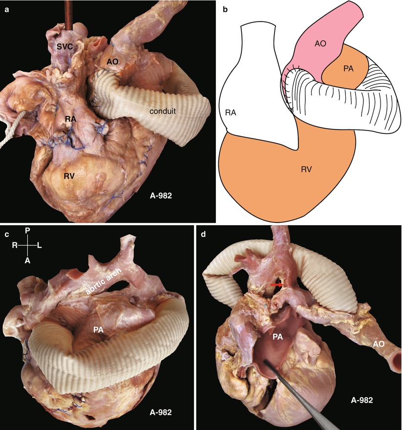
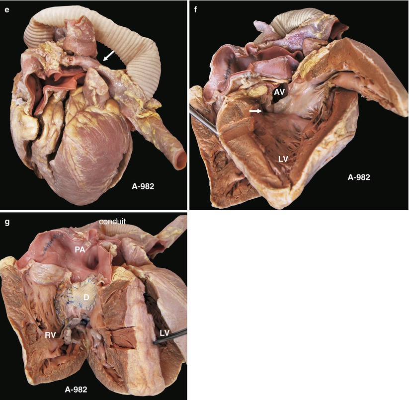
Congenital narrowing between the innominate artery and the left subclavian artery is called the aortic arch coarctation. Most of the aortic arch coarctation is located between the left common carotid artery and the left subclavian artery. The coarctation can be divided into a membrane-like stenosis or a tube-like constriction (Figs. 15.6, 15.1, 15.2, 15.7, and 15.3).
15.3 Interrupted Aortic Arch
Interrupted aortic arch is defined as luminal atresia or discontinuity between the ascending aorta and the descending aorta. Some have a residual aortic wall structure. They are categorized into three types, type A, type B, and type C, according to the site of the interruption. The three types are further classified into several subtypes based on the opening and closure of the ductus arteriosus and the concurrent cardiac malformations.
15.3.1 Type A
In type A, the site of the interruption is located between the left subclavian artery and the patent ductus arteriosus. When a concurrent VSD is present, the descending aorta receives the mixed blood from the pulmonary artery and oxygenated blood from the ascending aorta through collateral circulation. Infants with this condition may survive to childhood, usually with pulmonary hypertension. The goals of surgical correction of type A interrupted aortic arch are (1) to build communication between the ascending aorta and the descending aorta and (2) to cut off the connection between the patent ductus arteriosus and the pulmonary artery. Considering that the traditional method of dissecting the free patent ductus arteriosus and aorta involves a complicated procedure and a high risk of bleeding, we use the pulmonary intracavitary correction method without dissecting free the great vessels, which renders the repair of the VSD easier.
15.3.2 Type B
In the type B, the site of interruption is located between the left subclavian artery and the left common carotid artery.
15.4 Specimen Demonstrations
15.4.1 Aortic Arch Coarctation, Conduit Connected with the Aortic Arch and the Descending Aorta


Fig. 15.1
(a) Specimen 982. Aortic arch coarctation. The ascending aorta and the descending aorta are connected with the artificial blood vessel. The figure demonstrates the proximal anastomosis of the artificial blood vessel (conduit). AO aortic arch, RA right atrium, RV right ventricle, SVC superior vena cava. (b) Specimen 982. Schematic diagram of Fig. 15.1a. (c) Specimen 982. The ascending aorta and descending aorta viewed from the cephalic position. The aortic arch coarctation (aortic arch), ascending aorta, and descending aorta are connected with the artificial blood vessel. PA pulmonary artery. (d) Specimen 982. View from the anterior aspect of the heart. The arrow indicates the coarctation at the distal part of the left common carotid and the dilated main pulmonary artery (PA). (e) Specimen 982. Aortic arch coarctation viewed from another visual angle (arrow). (f) Specimen 982. View of ventricular septal defect (arrow) from the left ventricle. AV aortic valve, LV left ventricle. (g) Specimen 982. Aortic arch coarctation. The VSD patch (D) is viewed from the RV; the distal coarctation of the aortic arch, with concurrent VSD, is shown
< div class='tao-gold-member'>
Only gold members can continue reading. Log In or Register to continue
Stay updated, free articles. Join our Telegram channel

Full access? Get Clinical Tree


