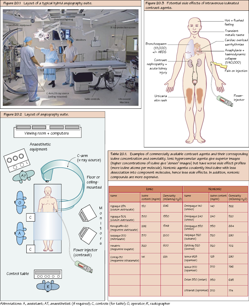
Angiography I: Overview Catheter-directed angiography is still the ‘gold standard’ in vascular imaging, but it is invasive (intra-arterial instrumentation) and therefore often reserved for those likely to require a simultaneous therapeutic intervention. Any stenosis identified (taken in at least two planes) is ‘percentage’ quantified according to the degree of luminal narrowing (50%, 75%, 90%, 100% [total occlusion]). This is a purpose-built room for diagnostic and therapeutic intervention often located in the radiology department, although in modern vascular practice the suite often doubles as an operating room (hybrid suite) located within the main theatre complex. The room must adhere to national radiation safety standards. Radiation exposure decreases proportionately by the square of the distance from the source.

The Angiography Suite
Personnel
Equipment
Basic Steps in Angiography
Radiation Safety
Stay updated, free articles. Join our Telegram channel

Full access? Get Clinical Tree


