Alveolar Pneumocytes, Reactive and Metaplastic Changes
Allen P. Burke, M.D.
Acute Reactive Changes
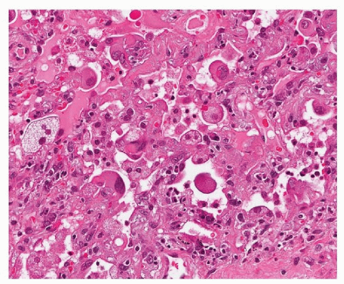
Alveolar lining cells are denuded and regenerative in acute alveolar injury. Histologic features of regeneration include nuclear enlargement, cytoplasmic enlargement, and cytoplasmic vacuolization. Extensive vacuolization that involves macrophages as well is typical of amiodarone toxicity and Hermansky-Pudlak syndrome. Reactive pneumocytes persist from acute to organizing stage of alveolar injury (Figs. 6.1, 6.2, 6.3). In some cases, the changes suggest viral cytopathic effects.
Chronic Reactive Changes
With loss of fibrin and transformation of the interstitium to a more restive, fibrotic state, type II pneumocytes are less atypical but maintain a crowded configuration with cuboidal shape. In this phase, they are most difficult to distinguish from preneoplastic changes, such as atypical adenomatous hyperplasia (AAH). AAH tends to be relatively circumscribed and uniform, whereas reactive changes are more diffuse and show areas typical of reactive fibrosis (Figs. 6.4, 6.5, 6.6, 6.7).
Mucinous and Squamous Metaplasia
Squamous metaplasia is a reactive feature in both acute lung injury, such as diffuse alveolar damage and acute respiratory distress syndrome, as well as chronic lung injury (Fig. 6.8). In the latter, it is typically seen in the variegated lining of remodeled cystic cavities in usual interstitial pneumonia.
 FIGURE 6.1 ▲ Acute pneumocyte hyperplasia, acute radiation pneumonitis. A variety of acute lung injury can result in reactive atypia in regenerating pneumocytes. The lining cells are markedly enlarged with vacuolated cytoplasm and prominent nucleoli. In addition, there is inflammation in the thickened alveolar septa.
Stay updated, free articles. Join our Telegram channel
Full access? Get Clinical Tree
 Get Clinical Tree app for offline access

|