Alternative Cardiac Imaging Techniques
Echo is not the only technique for obtaining diagnostic information about the heart. Several cardiac imaging techniques are available, some of which provide similar information to echo (e.g. assessment of left ventricular (LV) function with cardiac magnetic resonance imaging (MRI)) and some of which can provide additional information about the heart that echo alone cannot obtain (e.g. visualization of coronary artery stenoses with coronary angiography). Knowledge of these alternative imaging techniques can help you decide when another test might replace or supplement an echo study.
NUCLEAR CARDIOLOGY
Nuclear cardiology uses radioactive isotopes, administered intravenously, to image the heart and to provide information about myocardial perfusion (myocardial perfusion imaging) and ventricular function (radionuclide ventriculography).
Uses of nuclear cardiology
Myocardial perfusion imaging uses a radiopharmaceutical (e.g. thallium-201 or a technetium-99m-labelled radiopharmaceutical) to assess myocardial blood flow, providing valuable information about coronary artery disease with a high degree of sensitivity and specificity (Fig. 14.1).
After the radiopharmaceutical has been administered intravenously, its distribution in the myocardium can be assessed using single photon emission computed tomography (SPECT) imaging. Imaging is performed at rest and again after stress (exercise or pharmacological), and comparison of the rest and stress images allows identification of areas of normal perfusion, reversible ischaemia (normal perfusion at rest but reduced perfusion after stress) and fixed ischaemia (reduced perfusion at rest and after stress). The use of ECG gating also allows myocardial function to be assessed with the calculation of a LV ejection fraction.
Radionuclide ventriculography gives an accurate assessment of ventricular function. It is most commonly performed using red blood cells labelled with technetium-99m which are administered intravenously. The count-rate of the radioactivity can be measured using a gamma camera over many cardiac cycles and, with the use of ECG gating, the average count rate at different stages of the cardiac cycle can be calculated. From this, an accurate measure of ejection fraction can be derived.
DISADVANTAGES OF NUCLEAR CARDIOLOGY
The principal drawback of nuclear cardiology is the patient’s exposure to ionizing radiation. The typical effective radiation dose of a 99mTc dynamic cardiac scan is 6 mSv, equivalent to 2.7 years’ exposure to natural background radiation. For a
201Tl myocardial perfusion scan the typical dose is 18 mSv, equivalent to 8 years’ background radiation (for comparison, the typical dose of a single chest X-ray is 0.02 mSv, or 3 days’ background radiation). Further information on radiation doses and the lifetime additional risk of fatal cancer can be found on the website of the Health Protection Agency (www.hpa.org.uk).
201Tl myocardial perfusion scan the typical dose is 18 mSv, equivalent to 8 years’ background radiation (for comparison, the typical dose of a single chest X-ray is 0.02 mSv, or 3 days’ background radiation). Further information on radiation doses and the lifetime additional risk of fatal cancer can be found on the website of the Health Protection Agency (www.hpa.org.uk).
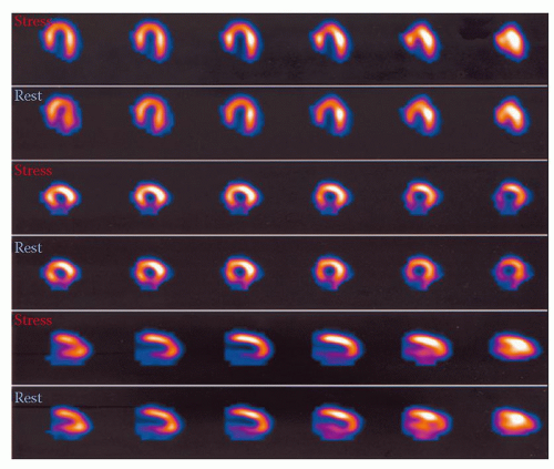 Figure 14.1 Myocardial perfusion imaging (showing inferior wall defect) |
CARDIAC MAGNETIC RESONANCE IMAGING
MRI is a highly versatile technique for cardiac imaging and provides both anatomical and functional information. Cardiac MRI is performed with a scanner containing a large superconducting magnet; radio waves are transmitted into the heart, aligning hydrogen nuclei, and as the nuclei subsequently ‘relax’ they emit radio waves of their own which can be detected by the scanner. The detected signal can then be used to reconstruct an image of the heart (Fig. 14.2).
Uses of cardiac MRI
Stay updated, free articles. Join our Telegram channel

Full access? Get Clinical Tree


