Contrast Echo
There are two very different types of echo contrast study, using:
agitated saline bubble contrast
echo contrast agents.
AGITATED SALINE BUBBLE CONTRAST
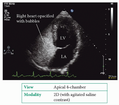
An agitated saline bubble contrast is simple to perform and is used primarily to detect a right-to-left shunt, most commonly a patent foramen ovale (PFO, p. 282). A suspension of tiny air bubbles is injected intravenously while an echo is performed. Normally the bubbles fill the right heart where they are clearly visible on echo (Fig. 9.1), but they are then filtered out as they pass through the lungs – no bubbles will therefore be seen in the left heart. If bubbles are seen within the left heart, this indicates that the agitated saline bubble contrast (and hence blood) is managing to cross directly from the right heart into the left via a right-to-left shunt, bypassing the lungs.
Normally the presence of an intracardiac shunt will allow blood to flow from left to right (high pressure to low pressure), but during a Valsalva manoeuvre the blood flow will momentarily reverse from right to left. To demonstrate a transient rightto-left shunt you can use ‘agitated’ saline:
Draw up 8.5 mL of normal saline and 0.5 mL of air into a 10 mL Luer lock syringe.
Using a 3-way tap, connect this to another (empty) 10 mL Luer lock syringe, and then attach this to an intravenous cannula sited in the patient’s antecubital vein.
Withdraw 1 mL of the patient’s blood into the syringe containing the saline/air mixture.
With the 3-way tap turned off to the patient, repeatedly squirt the saline/blood/air mixture back and forth between the two syringes for a few seconds until a suspension of tiny air bubbles is created in the mixture.
Obtain a good apical 4-chamber view with your echo probe.
With the patient performing a Valsalva manoeuvre, ensure you are recording the echo images and rapidly inject the 10 mL mixture. When the bubbles appear in the right atrium, ask the patient to release the Valsalva manoeuvre.
Watch carefully for air bubbles crossing into the left atrium as the patient releases the Valsalva – some authorities regard the crossing of a single bubble as indicative of a shunt, others require three or more bubbles before making the diagnosis.
An agitated saline contrast study can be performed during transthoracic (TTE) or transoesophageal (TOE) echo. Although the image quality is better with TOE, patients usually perform a better Valsalva manoeuvre during TTE.
An additional use of agitated saline bubble contrast is to assist with echo guidance during pericardiocentesis. If there is doubt about whether the pericardiocentesis needle is in the pericardial cavity, a small amount of agitated saline can be injected
through the pericardiocentesis needle. If the needle is in the right place, the bubbles will appear within the pericardial effusion. If the pericardiocentesis needle has inadvertently punctured the heart, the bubbles will be seen within one of the cardiac chambers instead.
through the pericardiocentesis needle. If the needle is in the right place, the bubbles will appear within the pericardial effusion. If the pericardiocentesis needle has inadvertently punctured the heart, the bubbles will be seen within one of the cardiac chambers instead.


