Air-Containing Mediastinal Mass
Toms Franquet, MD, PhD
DIFFERENTIAL DIAGNOSIS
Common
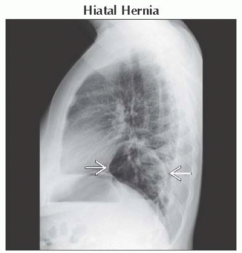
Hiatal Hernia
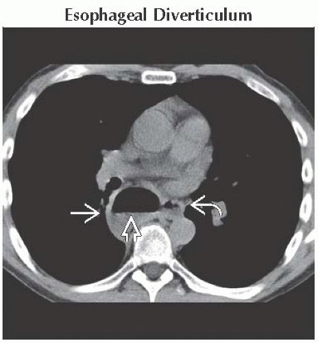
Esophageal Diverticulum
Zenker Diverticulum
Less Common
Achalasia
Esophageal Perforation
Mediastinal Abscess
Rare but Important
Bronchogenic Cyst
Loculated Pneumomediastinum
ESSENTIAL INFORMATION
Key Differential Diagnosis Issues
Location of air is key
Retrocardiac mass with air or air-fluid level is characteristic of esophageal hiatus hernia
Superior mediastinal air-fluid level often Zenker diverticulum
Bronchogenic cyst usually subcarinal
Helpful Clues for Common Diagnoses
Hiatal Hernia
± air-fluid level
Esophageal Diverticulum
Air-fluid level in mid-esophagus (traction) or in epiphrenic region (pulsion)
Zenker Diverticulum
Air-fluid level in superior mediastinum (pharyngoesophageal junction)
Complications: Aspiration pneumonia and bronchiolitis
Helpful Clues for Less Common Diagnoses
Achalasia
Esophageal dilatation with air-fluid level
Esophageal Perforation
Penetrating trauma (90%): Iatrogenic following endoscopic procedures, postsurgical and ingested foreign bodies (e.g., bones)
Spontaneous: Boerhaave syndrome (esophageal rupture after emesis)
Radiographic findings: Pneumomediastinum and subcutaneous emphysema in soft tissues of neck
NECT: Gas collections centered around esophagus (90%)
Mediastinal Abscess
Usually secondary to erosion from esophageal carcinoma
Following cardiac or esophageal surgery
Helpful Clues for Rare Diagnoses
Bronchogenic Cyst
Spontaneous cyst ruptures into airways, esophagus, pleural cavity, and pericardial cavity
Radiograph & CT: Large subcarinal cyst with air-fluid level (rupture)
Loculated Pneumomediastinum
In neonates, air in mediastinum often loculates locally; tends not to dissect widely as in adults
Image Gallery
 Axial NECT shows esophageal diverticulum
 with a visible air-fluid level with a visible air-fluid level  . Adjacent esophagus is also seen . Adjacent esophagus is also seen  . This is consistent with a pulsion esophageal diverticulum. . This is consistent with a pulsion esophageal diverticulum.Stay updated, free articles. Join our Telegram channel
Full access? Get Clinical Tree
 Get Clinical Tree app for offline access

|

