Additional Considerations for the Endovascular Treatment of Extracranial Carotid Artery Occlusive Disease


Additional Considerations for the Endovascular Treatment of Extracranial Carotid Artery Occlusive Disease


Peter A. Schneider


The natural history, principles of management, and operative techniques for both open and endovascular revascularization of extracranial carotid artery occlusive disease have been described in the preceding chapters, and the following two chapters will cover recurrent stenosis and carotid body tumors. In this author’s opinion, the most important additional consideration with respect to managing carotid artery occlusive disease is the upcoming transition from endarterectomy to carotid angioplasty and stenting (CAS) as the primary treatment modality. In preparation for the transition, the following issues will be addressed in this chapter: the requisite skills necessary for CAS; the indications for carotid arteriography in the era of CAS; the technique of carotid arteriography; and the technical aspects of cerebral protection devices.


Requisite Skills for Carotid Angioplasty and Stenting

Vascular surgeons are well versed in the natural history, clinical evaluation, surgical management, noninvasive assessment, and follow up of extracranial carotid artery occlusive disease. Indeed, surgeons may know too much; familiarity with the lumen of the atherosclerotic carotid bifurcation has made us hesitant to believe that CAS could be safe or effective. The endovascular skills acquired in other vascular beds may be transferred to the carotid system with some caveats (Table 29-1). Surgeons must develop the necessary tools/skills to assess the aortic arch, catheterize the carotid arteries, pass sheaths from a remote access, and use the rapid exchange or monorail systems. With the development of carotid duplex and the other noninvasive imaging modalities, fewer arteriograms (i.e., arch, carotid, cerebral) have been performed for carotid occlusive disease over the last 10 years. Future therapy will depend upon an understanding of the arch and its anatomical features. It is essential that surgeons be facile with carotid arteriography.


Aortic Arch Assessment

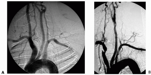
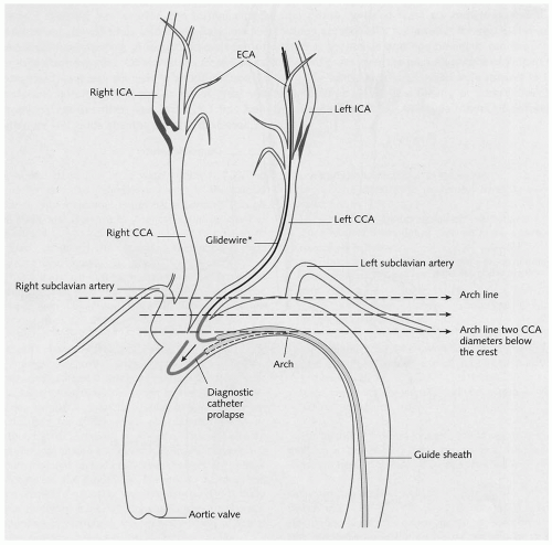
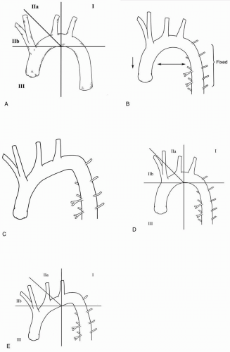
Most methods of assessing the aortic arch are designed to take its general shape into account. The arch tends to elongate in association with age and long-standing hypertension. Because the proximal descending aorta is relatively “fixed” by the mediastinum and intercostal arteries, the arch tends to assume a sloping configuration with the aortic valve slightly depressed in the chest and the distal arch coming to a peak before turning caudally (Fig. 29-1). One frequently used classification system involves drawing a horizontal line across the “top” of the arch. If the branches originate at the “top” of the arch, it is classified as a level 1. If the branches originate a distance of one or two common carotid artery diameters caudal to this line, it is referred to as a level 2 or 3 arch, respectively (Fig. 29-2). The more the arch tends to slope and the farther caudal along the slope that the branches originate, the more challenging they are to catheterize and the more difficult it is to pass a sheath. However, when carotid stent placement is the goal, only one common carotid artery requires sheath access, and it is the location of this target artery that is relevant. In addition, the location of the “top” of the arch is of little functional consequence to the surgeon. The factor that most significantly determines the degree of difficulty for endovascular procedures is the location of the fulcrum of the arch (its upper inner aspect) relative to the branch vessels. Specifically, once the catheter or sheath passes over the fulcrum, the trajectory from the fulcrum to the target artery determines the level of difficulty. The “surf and turf” classification takes these anatomic factors into account (Fig. 29-3). A horizontal line is drawn across the peak of the inner curve of the arch, and a vertical line is drawn at the location where the arch peaks superiorly. An additional line bisects the angle formed between these horizontal and vertical lines, thereby dividing the arch segments into I, IIa, IIb, and III regions. Vessels originating in the segment III are the most challenging to cannulate.


Catheterization of the Carotid Arteries

Catheterization of the carotid arteries requires an understanding of both simple and complex curve catheters. Almost all segment I and segment II arteries may be catheterized with simple curve catheters. The more toward segment Ill the artery branch is located, the more likely that a complex curve catheter will be required. If a complex curve catheter is required, the secondary curve is usually seeded at the junction of the aortic arch and the common carotid artery, and the selective arteriogram is
performed with the catheter in this position. If the case proceeds to carotid stent placement, the elbow or secondary curve of the catheter must be advanced into the artery at its origin to obtain exchange guidewire access to the common carotid artery. Carotid artery catheterization is discussed in more detail below.


Table 29-1 Skills Required for Carotid Angioplasty and Stenting


Assessment and negotiation of the aortic arch


Selective catheterization of the carotid arteries


Carotid and cerebral arteriography


Passage of a sheath into a remote access


Understanding of rapid exchange or monorail systems


Technical aspects of cerebral protection devices


Passage of the Carotid Guiding Sheath

Working from a remote access site (i.e., femoral access for carotid interventions) poses several challenges (Table 29-2). Because the sheath courses over a long distance, redundancy may build up within it that can make subsequent movements difficult (less predictable) and catheter exchanges challenging. Experience with remote access site interventions can be obtained using a contralateral femoral approach for infrageniculate interventions and a brachial approach for renal or iliac interventions. Remote access work relies on the inner guidewire or catheter for support. If there is a lot of tortuosity, the sheath may “fall” into the aortic arch when the guidewires or catheters are withdrawn. The presence of the sheath tip in the common carotid artery may accentuate or correct the tortuosity in the common or internal carotid arteries, depending upon its angle of approach. During carotid bifurcation stenting, the sheath tip is advanced to the mid-common carotid artery so that the tip of the sheath and the cerebral protection device (positioned in the distal internal carotid artery) may be included in the field of view. The sheath must be advanced far enough into the artery so that it is well anchored, and this is particularly relevant after the exchange catheter is removed. Care is taken to avoid mechanical dilatation of the lesion by the tip of the dilator during sheath advancement.

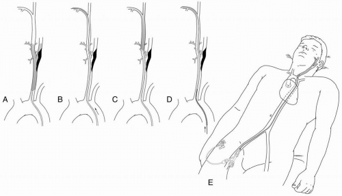
After common carotid artery catheterization with the selective catheter, the image intensifier is placed in a position that “opens” or splays out the carotid bifurcation (Fig. 29-4). This angle may vary significantly from one patient to the next but is usually somewhere between a straight lateral and a steep oblique. The carotid bifurcation is roadmapped, and a 260 cm length, 0.035 in Glidewire (Boston Scientific, Natick, Mass.) is advanced into the external carotid artery. The selective catheter is advanced into the external carotid artery and the guidewire is removed. The tip of the catheter must be advanced a few cm inside the external carotid artery so that it does
not become inadvertently dislodged into the carotid bulb with breathing or arterial pulsation. The external carotid artery is roadmapped, and the best branch for anchoring the carotid sheath is chosen. The guidewire is advanced into the distal external carotid artery branch and is followed by the catheter. The Glidewire is subsequently removed. The selective cerebral catheter must be back bled to avoid introducing air into the system. This often requires withdrawing the catheter slightly, because its tip usually enters a small, distal branch. The exchange guidewire is then placed in the external carotid artery. Commonly used exchange guidewires are the Amplatz super-stiff (Cook, Inc., Bloomington, IN) or extra-stiff (Boston Scientific), the Supracore (Guidant, Menlo Park, CA), the Microvena Nitinol (Microvena Corp., White Bear Lake, MN), or a Stiff Glidewire (Boston Scientific). A braided, selective catheter is useful when advancing the stiff exchange guidewire into the external carotid artery, because it is less likely to be pulled out. After the exchange guidewire has been placed, the selective catheter is removed and the long, carotid guiding sheath is inserted. The guidewire is surveyed with fluoroscopy to look for any redundant segments; these should be removed before sheath passage. The 6 Fr sheath is commonly used with popular ones, including the Shuttle Sheath (Cook), Destination (Boston Scientific), and Vista Brite Tip (Cordis Corp., Miami Lakes, FL). The sheath is advanced over the exchange guidewire with steady forward pressure. The field of view should include the guidewire tip and the course of the guidewire from the arch into the common carotid artery to make certain that the guidewire is not migrating caudally. When the sheath tip reaches the last major turn from the arch into the common carotid artery, the angle of approach can be made less acute by having the patient take a deep breath.


Figure 29-1. Normal versus elongated arch. A: This configuration of the aortic arch is relatively “normal.” B: This arch aortogram demonstrates elongation of the ascending and transverse aortic segments in an elderly hypertensive man. The junction of the distal arch and the proximal descending aorta comes to a peak where the aorta is “fixed” in the posterior mediastinum. The arch branches originate along its “up slope.”


Figure 29-2. The diagram shows the designation of the arch levels 1, 2, or 3. A horizontal line is drawn across the “top” of the arch. The level 1 arch has branches originating along that line, from the “top” of the arch. The level 2 arch has branches originating more than one common carotid artery diameter caudal to the “top” of the arch. The level 3 arch has its branches originating more than two common carotid artery diameters caudal to the “top” of the arch. (Reproduced with permission from Myla S. Carotid access techniques: an algorithmic approach. Carotid Interv. 2001;3:2-12.)


Figure 29-3. Aortic arch classification using the “surf and turf” classification. A: A horizontal line is drawn across the peak of the inner curve of the arch. This point forms a fulcrum and is the location over which the catheters must work to achieve carotid access. A vertical line is drawn at the location where the arch peaks superiorly. An additional line bisects the angle formed between these horizontal and vertical lines, thereby dividing the arch segments into I, IIa, IIb, and III regions. The further caudally and toward the patient’s right-hand side the branch vessels originate, the more challenging they are to catheterize and achieve sheath access. B: Normal arch. C: Elongated arch that “pushes” the origins of the branch vessels down into the chest and causes more acute curvature of the distal arch. This is representative of elderly patients with long-standing hypertension. D: When the “surf and turf” classification is applied to the normal arch, the left common carotid artery is in segment IIa, and the innominate and right common carotid artery are in segment IIb. E: The “surf and turf” classification applied to the elongated arch shows that the innominate and right common carotid arteries originate in segment III. (Reproduced with permission from Schneider PA. Carotid arteriography. In: Schneider PA, Bohannon WT, Silva MB Jr, eds. Carotid Interventions. New York: Marcel Dekker Inc, 2004:36.)


Rapid Exchange or Monorail Systems

Rapid exchange or monorail systems, especially using the low-profile 0.014-in system, are the likely platforms for all carotid interventions in the future. Notably, the distal filters and occlusion balloons used for cerebral protection are on 0.014-in platforms. The guidewire lumen extends only a short distance (i.e., 30 cm along a 130-cm length catheter) in the monorail system rather than along the entire length as with the coaxial systems (Table 29-3). The advantages of this system are the decrease in friction associated with passing the catheters
and the lower profile. The main disadvantage of the monorail system is that it must be delivered directly into the side branch through a long sheath (rather than over a large-caliber guidewire). There is a learning curve associated with its use, but experienced vascular specialists consider it easier and faster than the standard coaxial system.


Table 29-2 Pitfalls Associated with Remote Access and Working Through a Long Sheath


Must have an adequate length of exchange guidewire in the target vascular bed in order to pass a long sheath.


As the sheath is passed into the carotid artery, must be aware of the location of the sheath tip so that it doesn’t inadvertently dilate the lesion.


An adequate length of sheath tip must be in the target vascular bed to prevent it from collapsing into the aortic arch.


Once the dilator is removed, it is usually not possible to advance the sheath. The sheath may be withdrawn, but it may jump back a greater distance than desired.


Redundancy may accumulate within the sheath. This redundancy must be removed if possible.


Redundancy tends to accentuate curves and cause kinks that can become an obstacle to passing stents and other devices through the sheath.


Must observe the access sheath using fluoroscopy as it is being placed. If it meets an obstacle, it may kink or cause arterial injury.


Must observe the access sheath using fluoroscopy when removing the exchange guidewire, because the sheath may buckle or migrate inferiorly when the supporting wire is removed.


Without the stiff inner guidewire in place, the sheath configuration usually changes.


Indications for Carotid Arteriography

Carotid arteriography has been used selectively before carotid endarterectomy in institutions with reliable carotid duplex studies. The use of carotid duplex as the sole study before carotid repair is based on two principles:


  • The refinement of its accuracy to determine the degree of stenosis


  • The benefits of the arteriogram, in terms of reducing the complication rates of the open repair, do not outweigh its small risk

The advent of CAS has expanded the role of carotid arteriography, which currently serves as the best imaging study to select patients for CAS and should be performed routinely. It is important to emphasize that the resurgence of carotid arteriography is not based upon a desire to determine the degree of stenosis, because the noninvasive imaging is adequate for this purpose. The expanding role for carotid arteriography has been supported by its improved safety and the importance of knowing the status and configuration of the arterial tree from the arch to the intracranial portion of the internal carotid before CAS. Carotid arteriography includes many of the same key steps as CAS, and patients that cannot tolerate carotid arteriography cannot be treated with CAS. Selective use of carotid arteriography is still reasonable in patients who are candidates for carotid endarterectomy. The current indications for carotid arteriography are listed in Table 29-4. Vascular specialists must be facile with carotid arteriography to participate in the management of carotid artery occlusive disease.


Figure 29-4. Placement of the sheath is illustrated. A: The common carotid artery is catheterized with a selective catheter, and a roadmap of the carotid bifurcation is obtained with the image intensifier rotated in such a way that the bifurcation is “opened up” or splayed out. B: A steerable guidewire is advanced into the external carotid artery, and the selective catheter is advanced over it. C: The selective catheter is used to perform a roadmap of the external carotid artery, and a long branch of the external carotid artery is chosen to anchor the stiff guidewire. D: The selective catheter is advanced into the distal segment of the external carotid artery branch, and a stiff exchange guidewire is placed. E: The short femoral access sheath is removed, and a long carotid guiding sheath is placed with its tip in the common carotid artery. (Reproduced with permission from Schneider PA. Access for carotid interventions. In: Schneider PA, Bohannon WT, Silva MB Jr, eds. Carotid Interventions. New York: Marcel Dekker Inc, 2004:100-102.)


Technique of Carotid Arteriography


Access and Supplies for Carotid Arteriography

The first choice for access is the femoral artery (either side). If femoral access is contraindicated, the left brachial artery should be used. A towel is placed on the patient with the supplies required for access: local anesthetic, a scalpel, a clamp, a puncture needle, a guidewire, and a 4 or 5 Fr sheath. The inguinal ligament is traced from the anterior superior iliac spine to the pubic tubercle, and the proximal femoral artery is located. After anesthetic infiltration of the skin and subcutaneous tissue, the common femoral artery is punctured with the needle at a 45° angle of approach. The dominant hand advances a floppy tip and starts the guidewire through the needle. Fluoroscopy is used to monitor the advancement of the guidewire into the abdominal aorta. Use of a hemostatic access sheath for carotid arteriography is advisable. The sheath simplifies the catheter exchanges and reduces the friction at the access site that can adversely affect catheter rotation and advancement during selective carotid catheterization. A 4 or 5 Fr sheath can usually be placed over a starting guidewire. The sidearm port of the
sheath is directed toward the surgeon, and pressure is maintained at the arteriotomy with the nondominant hand until the sheath enters the artery.


Table 29-3 Comparison of Coaxial and Monorail Systems


Coaxial


Monorail


Long guidewire lumen along catheter


Guidewire lumen is shorter


Higher friction of catheter over guidewire


Lower friction; guidewire lumen is short


Higher profile


Lower profile, usually a 0.014-in system


Delivery with large caliber guidewire


Delivery with guiding sheath


Longer guidewire required


Shorter guidewire satisfactory


Supplies required for carotid arteriography are listed in Table 29-5. A 180-cm length, 0.035-in general purpose guidewire with a floppy tip (e.g., Bentson) is advanced into the arch of the aorta. A 90- or 100-cm long, 4 or 5 Fr flush catheter (e.g., pigtail) is placed under fluoroscopic guidance, and the arch aortogram is performed. A selective cerebral catheter is chosen based upon the configuration of the aortic arch. The flush catheter is exchanged for a selective catheter over a hydrophilic, steerable 260-cm long, 0.035-in guidewire (e.g., Glidewire, Boston Scientific). Selective cerebral catheters have a diameter of 4 or 5 Fr, a single end hole, and a specially shaped catheter tip, and are 90 to 125 cm in length. The selective catheter is advanced into the arch branch over the steerable guidewire.


Handling of Guidewires and Catheters

Excellent guidewire and catheter hygiene is imperative during all carotid arteriography to prevent the development of thrombus and embolization. All guidewires are wiped with a heparinized saline solution before placement and after removal. Similarly, catheters are flushed and wiped before insertion and after each exchange. After a catheter is placed, it should be gently aspirated and flushed while maintaining the syringe in a vertical position to trap any air bubbles. Catheters are flushed sparingly after insertion. Caution must be exercised to assure that no unintended solutions are infused. When connecting the catheter to extension tubing for the power injector, avoid leaving air bubbles in the line. Guidewires should be withdrawn from the catheters slowly and without whipping them to avoid creating suction in the catheter (and potential air emboli).


Table 29-4 Current Indications for Carotid Arteriography for Extracranial Carotid Artery Occlusive Disease


Only gold members can continue reading. Log In or Register to continue

Stay updated, free articles. Join our Telegram channel

Jun 16, 2016 | Posted by in CARDIAC SURGERY | Comments Off on Additional Considerations for the Endovascular Treatment of Extracranial Carotid Artery Occlusive Disease

Full access? Get Clinical Tree

Get Clinical Tree app for offline access

History


Physical Exam


Carotid Duplex


Treatment