Acute Pericarditis/Pericardial Effusion


Acute Pericarditis/Pericardial Effusion


Elina Yamada

Mani A. Vannan

Mauro Moscucci


ACUTE PERICARDITIS


Definition

The pericardium is a double-layered wrapping around the heart, with the outer and inner layers called parietal and visceral pericardium, respectively. The cavity between these two layers usually contains a small amount (15 to 50 mL) of an ultrafiltrate of plasma, which serves to lubricate the surfaces. Clinically relevant disorders of pericardium are acute inflammation (acute pericarditis), pericardial effusion with or without hemodynamic compromise (tamponade), constrictive pericarditis, congenital absence of the pericardium, and pericardial cysts.

Acute pericarditis is a condition that is first seen with the following clinical signs and symptoms: chest pain, pericardial friction rub, and typical electrocardiographic (ECG) changes. At least two of these features must be present to support the diagnosis.


Usual Causes

A wide variety of conditions can affect the pericardium, as listed in Table 26.1. The yield of the standard diagnostic evaluation is very low, about 16%. Idiopathic post viral, neoplastic, connective tissue disorders and uremia account for most acute pericarditis diagnosed in the clinical setting. When upper respiratory symptoms precede acute cardiac involvement, the most common etiology is post viral pericarditis. Coxsackie A or B virus or echovirus are the common agents (1). The term acute idiopathic pericarditis applies when there is no clear etiology identified and it is presumed to be viral or autoimmune. Viral serologic testing has a very low diagnostic yield and does not change management. Therefore it is not routinely recommended. Among the entities listed in Table 26.1, the human immunodeficiency virus (HIV) is an increasingly common etiology for the acute pericarditis (2,3) that is the most frequent cardiovascular manifestation of AIDS. The condition may be caused by HIV itself or may result from opportunistic infections or neoplasms (such as lymphoma). The presence of pericardial effusion in HIV syndrome is associated with poor prognosis.

Acute pericarditis should be considered in the differential diagnosis in the presence of hemodynamic deterioration after cardiac procedures or with new radiographic cardiomegaly.

Symptoms

A cardinal symptom of acute pericarditis is chest pain. The typical pain of pericardial inflammation is a retrosternal sharp pain radiating to the back near the trapezius edge. It is usually worse when the patient is in a supine position, and it is either relieved or ameliorated by sitting up. Chest pain may, however, be variable in location, nature, intensity, and radiation. It can be located retrosternally and radiate to the left arm, mimicking ischemic cardiac chest pain. It
may radiate to the epigastrium, mimicking abdominal disease, or worsen on deep inspiration, mimicking pleural pain. Constitutional symptoms are nonspecific and may include dyspnea, general malaise, weakness, hiccups, and cough. A low-grade fever may be present, but occasionally the body temperature may be as high as 40°C.


TABLE 26.1. Causes of acute pericarditis


Idiopathic


Infectious


Bacterial


Viral


Mycobacterial


Fungal


Protozoal


AIDS associated


Neoplastic


Primary


Secondary (breast, lung, melanoma, lymphoma, leukemia)


Immune inflammatory


Connective tissue diseases (rheumatoid arthritis, systemic lupus erythematosus, scleroderma, acute rheumatic fever, mixed connective tissue disease, Wegener granulomatosis) Arteritis (temporal arteritis, polyarteritis nodosa, Takayasu arteritis)


Acute myocardial infarction (MI) and post-MI (Dressler syndrome)


Postcardiotomy


Posttraumatic


Metabolic


Nephrogenic


Aortic dissection


Myxedema


Amyloidosis


Iatrogenic


Radiation injury


Procedures (cardiac catheterization, implantable defibrillator, pacemakers catheters, ablation)


Drugs (hydralazine, procainamide, daunorubicin, isoniazid, anticoagulants, cyclosporine, methysergide, phenytoin, dantrolene, mesalazine)


Cardiac resuscitation


Traumatic


Blunt trauma


Penetrating trauma


Surgical trauma


Congenital


Pericardial cysts


Congenital absence of pericardium


Mulibrey nanism syndrome


AIDS, acquired immunodeficiency syndrome.


Reproduced with permission from: Restivo J, Hoit B. Manual of Cardiology. New York: McGraw-Hill.


Signs

Tachycardia and tachypnea are usually nonspecific signs reflecting the general syndrome. However, they may indicate myocardial inflammation or hemodynamic compromise or both. The presence of a pericardial rub is the most specific sign of acute pericarditis. It is a squeaky or scratchy sound best heard at the left lower parasternal border with the diaphragm of the stethoscope (4). It may be present in one (15%), two (33%) or three phases (56%) of the cardiac cycle, and the intensity of the sound may change with position and respiration. Furthermore, it may be present or absent within a given day. Suspension of respiration may allow distinction from a coexistent pleural friction rub.


Helpful Tests


Electrocardiogram

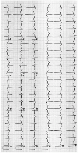
The ECG usually shows normal sinus rhythm except in the case of complicating arrhythmias. Atrial arrhythmias, when present, are usually associated with concomitant myocarditis or unrelated cardiac disease. Diffused ST-segment elevation and PR-segment depression may be present (Fig. 26.1), which then undergoes a typical evolutionary change, as listed in Table 26.2. These evolutionary changes in the ECG are pathognomonic of acute pericarditis, even in the absence of an audible pericardial friction rub (5,6). ECG changes always reflect a degree of myocardial involvement, inasmuch as the parietal pericardium is electrically inert. Bundle-branch block, intraventricular conduction delay, or Q waves may suggest myocardial involvement.


FIGURE 26.1. Electrocardiogram in a patient with acute pericarditis, showing diffuse ST-segment elevation and PR-segment depression.


TABLE 26.2. Electrocardiographic changes in acute pericarditis


STAGE


TIME COURSE


ECG CHANGES


1


ST-segment elevation occurs within hours of onset of chest pain and may persist for days


Upward concave ST-segment elevations, usually not exceeding 5 mm; PR segment depression (except in aVR)


2


Hours to days after stage 1


ST segments return to baseline: T waves are normal or show loss of amplitude


3


T-wave inversions may persist indefinitely (especially when associated with TB, uremia, or neoplasm)


T-wave inversions


4


Usually completed within 2 wk, but variability common


ECG normalizes


ECG, electrocardiographic; TB, tuberculosis.


Reproduced with permission from: Restivo J, Hoit B. Manual of Cardiology. New York: McGraw-Hill.


Early repolarization variant is in the differential of the acute ST-elevation pattern seen in pericarditis. Studies have shown that ST elevations usually occur in both limb and precordial leads in most cases of acute pericarditis, whereas about half of cases with the early repolarization variant have no ST changes in limb leads.


Chest Radiograph

The chest radiograph may be entirely normal or may show evidence of cardiomegaly when it is associated with pericardial effusion or myocarditis complicated by cardiac enlargement. In the setting of acute left ventricular failure, pulmonary congestion or signs of pulmonary edema may be seen.


TABLE 26.3. Echocardiography in pericardial effusion


Echolucent space between visceral and parietal layers of the pericardium


Typically does not extend beyond the left atrium and is anterior to the descending aorta in the parasternal long-axis view


Size and circumferential extent can be determined


Loculated effusions may be present after cardiac surgery, radiation, and infection


Partial organization and fibrin strands may be identified


RV early diastolic collapse and RA late diastolic collapse indicate elevated pericardial pressure (elevated RV pressures, as in pulmonary hypertension, may mask this sign)


Right-sided chambers collapse is not necessarily tamponade (PPV of 58%, NPV of 92%)


>40% and >25% peak-velocity respiratory variation in TV and MV Doppler flows may indicate hemodynamic compromise (obesity, COPD, LV dysfunction, and large pleural effusion can also cause respiratory variation)


Ascending aorta dissection flap may be detected as possible etiology of pericardial effusion


Echocardiography is helpful in guiding pericardiocentesis


COPD, chronic obstructive pulmonary disease; LV, left ventricular; MV, mitral valve; NPV, negative predictive value; PPV, positive predictive value; RA, right atrial; RV, right ventricular; TV, tricuspid valve.


Echocardiography

Although in acute idiopathic or postviral pericarditis, significant pericardial effusion occurs in only a minority of cases, some degree of effusion may be present in up to 60% of all cases of acute pericarditis. The 2003 Task Force of the American College of Cardiology, American Heart Association, and the American Society of Echocardiography gave a class I recommendation for echocardiography in cases in which pericardial disease is suspected. Echocardiography provides estimation of size and site of the effusion and comprehensive assessment of hemodynamic compromise caused by the pericardial effusion (Table 26.3 and Figs. 26.2, 26.3, 26.4). Left ventricular systolic function may be affected
with associated myocarditis, and echocardiography is helpful in estimating the degree of myocardial dysfunction. A surface or transesophageal echocardiography may detect a ruptured aortic dissection causing pericardial effusion, or a pericardial tumor. Furthermore, echocardiography is useful for detection of complications of pericarditis, such as constrictive pericarditis. Although echocardiography can diagnose constrictive physiology, it is not a reliable technique to assess pericardial thickening. Magnetic resonance imaging and computed tomography are superior techniques in detecting pericardial thickening (7). Repeated follow-up echocardiography is not routinely recommended in asymptomatic patients with known small pericardial effusion.

Only gold members can continue reading. Log In or Register to continue

Stay updated, free articles. Join our Telegram channel

Aug 18, 2016 | Posted by in CARDIOLOGY | Comments Off on Acute Pericarditis/Pericardial Effusion

Full access? Get Clinical Tree

Get Clinical Tree app for offline access