We report a case of focal myocarditis in a young boy mimicking acute ST-segment elevation MI. He presented with chest pain and the EKG changes were consistent with infero-laeral ST-segment elevation MI. Coronary angiogram revealed smooth arteries with no obstruction. Troponin was significantly elevated and the echocardiogram exhibited mildly impaired LV function with hypokinetic inferior and lateral walls. Subsequently performed cardiac magnetic resonance imaging confirmed the diagnosis of myocarditis by exhibiting classic features of delayed gadolinium enhancement in the epi and mid-myocardial regions of the lateral wall sparing the sub-endocardial region. This case exhibits the use of cardiac magnetic resonance imaging for diagnosis in such scenarios as often if the angiogram is normal other differential diagnosis are often speculated without actual evidence
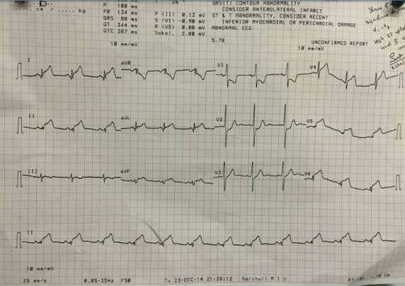
A 16-year-old boy with no relevant medical history presented to our institution with 3-hour history of nonradiating central crushing chest pain. There was no current or past use of cocaine or other stimulants. He was afebrile. His cardiovascular examination was unremarkable. The initial electrocardiography revealed sinus rhythm with inferolateral ST-segment elevation and ST-segment depression in the posterior leads ( Figure 1 ). Cardiac catheterization revealed smooth unobstructive epicardial coronary arteries ( Figure 2 , Supplementary Videos 1 to 3 ). He was given aspirin and intravenous morphine and his pain resolved. A repeat electrocardiography revealed persistent ST-segment elevation ( Figure 3 ). Initial troponin I was 48,979 ng/L (normal <26 ng/L). Echocardiography showed mildly impaired LV systolic function with hypokinetic inferior and lateral wall and no pericardial effusion. Cardiac magnetic resonance imaging (MRI) confirmed the diagnosis by demonstrating delayed gadolinium enhancement in the epi- and mid-myocardial regions of the lateral wall and with sparing of the subendocardial tissue, classic MRI features of myocarditis ( Figure 4 ). Patient made uneventful recovery and was discharged from hospital 48 hours later. He remained asymptomatic at 6-months’ follow-up with normalization of ST-segments changes ( Figure 5 ) and left ventricular function.