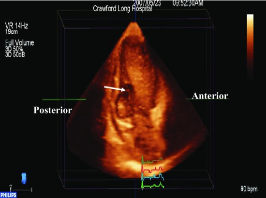
Figure 19.2 Three-dimensional transthoracic echocardiography: A circular clear area (arrow) in the inferior basal septum is noted in apical 2-chamber view that represents the septal perforation.
Surgical reduction of the aneurysm size and closure of the LV wall was carried out. The VSD was identified in the mid-septum posterior.
After the operation transthoracic echocardiography (TTE) demonstrated the LV size was normal and there was a small residual VSD.
Discussion
Stay updated, free articles. Join our Telegram channel

Full access? Get Clinical Tree


