Acute Embolic and Thrombotic Mesenteric Ischemia


Acute Embolic and Thrombotic Mesenteric Ischemia


Stuart I. Myers


Embolism and thrombosis of the mesenteric arteries, mesenteric venous thrombosis, and nonocclusive mesenteric ischemia are the four major causes of acute intestinal ischemia. Mortality rates for patients with acute mesenteric arterial occlusion range between 50% and 90%, and the presence and extent of bowel infarction predict mortality. A successful outcome depends on prompt diagnosis, rapid resuscitation, and surgical or endovascular revascularization, intestinal resection, and judicious use of a second-look celiotomy.


Clinical Presentation


Acute Embolic Mesenteric Ischemia


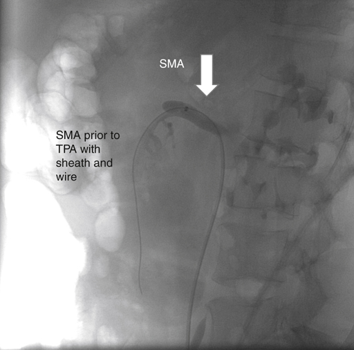
Acute embolic occlusion of the superior mesenteric artery (SMA) produces a characteristic syndrome of abdominal pain in 75% to 90% of patients and is often accompanied by vomiting and gastrointestinal emptying (Figure 1). A detailed history and physical examination usually elicit several important features. Patients might have no antecedent symptoms of chronic mesenteric ischemia, but between 30% and 43% of patients present with acute embolic occlusions superimposed on chronic occlusive disease. Approximately 75% of patients have a history of an atrial tachyarrhythmia, 30% have a history of embolic events, and 20% have synchronous emboli in other arterial beds. Atrial fibrillation, myocardial infarction, left ventricular aneurysm, prosthetic heart valves, and rheumatic heart disease can lead to the formation of thrombus that serves as a source of peripheral emboli. The classic finding of pain out of proportion to physical examination occurs because visceral ischemic pain precedes the somatic pain that is associated with transmural ischemia and peritoneal irritation. Peritoneal signs and sepsis are late findings associated with increasing mortality rates.


Several studies now report that acute emboli account for 28% to 42% of acute mesenteric ischemia. The common occurrence of acute emboli to the SMA may be a result of the oblique origin of this artery from the aorta. Fifteen percent of emboli lodge at the ostium of the SMA, 50% lodge distal to the proximal jejunal and middle colic branches, and 35% fragment and embolize distally. Embolic obstruction of the SMA produces ischemia from the level of the proximal jejunum to the transverse colon. The colon’s splenic flexure is often affected because it receives the terminal blood supply of both the SMA and inferior mesenteric artery (IMA) circulation.


Acute Thrombotic Mesenteric Ischemia


Acute thrombotic mesenteric arterial occlusion is now more common than embolic disease and ranges in incidence from 58% to 64%, has a different clinical presentation, and carries a similar high mortality rate. Patients are typically 60 years of age or older and have a history of symptomatic atherosclerotic occlusive disease affecting the carotid, coronary, or peripheral circulations. A history of intestinal angina, diarrhea, abdominal pain, and weight loss is present in up to 50% of patients who have acute mesenteric ischemia, yet the transition from chronic intestinal ischemia to the acute phase can present a diagnostic challenge. As in cases of acute mesenteric embolus, the patient’s complaints of abdominal pain are commonly out of proportion to the clinical findings. Patients with an antecedent history of chronic abdominal pain have often undergone extensive diagnostic testing and might even have had an operation that failed to relieve the pain.


Physical examination can reveal signs of malnutrition, and a detailed peripheral arterial examination often reveals absence of peripheral pulses and bruits in the distribution of the visceral or peripheral vessels. The abdominal examination may be unremarkable in the absence of peritoneal irritation, but 75% of patients will have occult blood in the stool.


Diagnosis


A shock state is present in approximately 25% of patients at initial evaluation, and this produces hemoconcentration, acidemia, and a leukocytosis that can exceed 20,000 white blood cells per millimeter. No single enzyme or combination is sensitive or specific enough to aid in the early diagnosis of intestinal ischemia. Serum hyperamylasemia is a nonspecific finding in 30% of patients with mesenteric ischemia. An elevated serum lactate is another nonspecific indicator of hypoperfusion with concomitant anaerobic metabolism.


Abdominal radiographs are usually nondiagnostic, and 25% of patients with mesenteric infarction have a normal abdominal radiograph. However, plain films can show thickened bowel loops, a ground-glass appearance caused by ascites, or a thumbprinting pattern caused by submucosal edema and hemorrhage. Free intraperitoneal air is present in patients with full-thickness bowel necrosis with perforation, and pneumatosis intestinalis and portal venous air also indicate intestinal infarction. Computed tomography (CT) can exclude other intraabdominal conditions that mimic intestinal ischemia, but CT scans are normal in up to 30% of patients with proven mesenteric ischemia. Duplex ultrasound and magnetic resonance imaging do not play important roles in the diagnosis of acute mesenteric ischemia.


Biplane arteriography allows early diagnosis and facilitates rapid surgical or endovascular treatment of patients with acute mesenteric arterial occlusions. Arteriography improves survival when it is performed before intestinal infarction and before surgical intervention. Arteriography allows one to differentiate nonocclusive from occlusive mesenteric ischemia and to institute immediate therapy to relieve mesenteric vasoconstriction and possibly the obstruction with endovascular techniques. Acute thromboses classically occur in the setting of advanced atherosclerotic occlusive disease and can involve the origins of all three primary mesenteric vessels, but embolic disease usually involves only the SMA origin with an abrupt cutoff of an otherwise normal-appearing artery.


Vasospasm contributes significantly to ongoing intestinal ischemia in both embolic and thrombotic occlusions. Thus the author begins a selective intraarterial papaverine infusion at 30 to 60 mg/hour once a diagnosis has been established. Boley, Feinstein, and Sammartano advocate continuation of the papaverine infusion until a repeat arteriogram documents resolution of the mesenteric vasoconstriction. Papaverine is a phosphodiesterase inhibitor that allows the accumulation of cyclic adenosine monophosphate (cAMP), with resultant vascular smooth muscle relaxation. Prostaglandin E1 and glucagon can also lead to the accumulation of cAMP and subsequent resolution of arterial vasospasm.

Stay updated, free articles. Join our Telegram channel

Aug 25, 2016 | Posted by in CARDIOLOGY | Comments Off on Acute Embolic and Thrombotic Mesenteric Ischemia

Full access? Get Clinical Tree

Get Clinical Tree app for offline access