Acute Abdomen

Chapter 47 Acute Abdomen


The term acute abdomen refers to signs and symptoms of abdominal pain and tenderness, a clinical presentation that often requires emergency surgical therapy. This challenging clinical scenario requires a thorough and expeditious workup to determine the need for operative intervention and initiate appropriate therapy. Many diseases, some of which are not surgical or even intra-abdominal,1 can produce acute abdominal pain and tenderness. Therefore, every attempt should be made to make a correct diagnosis so that the therapy selected, often a laparoscopy or laparotomy, is appropriate.


The diagnoses associated with an acute abdomen vary according to age and gender.2 Appendicitis is more common in younger individuals, whereas biliary disease, bowel obstruction, intestinal ischemia and infarction, and diverticulitis are more common in older adults. Most surgical diseases associated with an acute abdomen result from infection, obstruction, ischemia, or perforation.


Nonsurgical causes of an acute abdomen can be divided into three categories, endocrine and metabolic, hematologic, and toxins or drugs (Box 47-1).3 Endocrine and metabolic causes include uremia, diabetic crisis, addisonian crisis, acute intermittent porphyria, acute hyperlipoproteinemia, and hereditary Mediterranean fever. Hematologic disorders include sickle cell crisis, acute leukemia, and other blood dyscrasias. Toxins and drugs causing an acute abdomen include lead and other heavy metal toxins, narcotic withdrawal, and black widow spider poisoning. It is important to consider these possibilities when evaluating a patient with acute abdominal pain.


Because of the potential surgical nature of the acute abdomen, an expeditious workup is necessary (Box 47-2). The workup proceeds in the usual order—history, physical examination, laboratory tests, and imaging studies. Although imaging studies have increased the accuracy with which the correct diagnosis can be made, the most important part of the evaluation remains a thorough history and careful physical examination. Laboratory and imaging studies are usually needed, but are directed by the findings on history and physical examination.


Anatomy and Physiology


Abdominal pain is divided into visceral and parietal components. Visceral pain tends to be vague and poorly localized to the epigastrium, periumbilical region, or hypogastrium, depending on its origin from the primitive foregut, midgut, or hindgut (Fig. 47-1). It is usually the result of distention of a hollow viscus. Parietal pain corresponds to the segmental nerve roots innervating the peritoneum and tends to be sharper and better localized. Referred pain is pain perceived at a site distant from the source of stimulus. For example, irritation of the diaphragm may produce pain in the shoulder. Common referred pain sites and their accompanying sources are listed in Box 47-3. Determining whether the pain is visceral, parietal, or referred is important and can usually be done with a careful history.
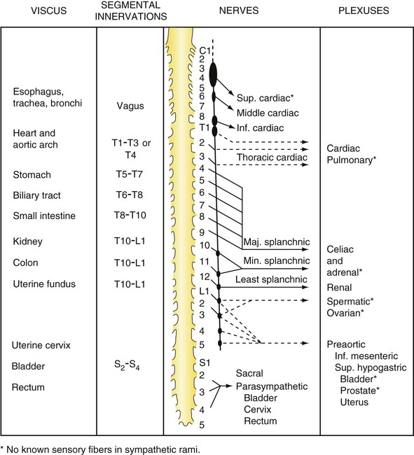


Introduction of bacteria or irritating chemicals into the peritoneal cavity can cause an outpouring of fluid from the peritoneal membrane. The peritoneum responds to inflammation by increased blood flow, increased permeability, and formation of a fibrinous exudate on its surface. The bowel also develops local or generalized paralysis. The fibrinous surface and decreased intestinal movement cause adherence between the bowel and omentum or abdominal wall and help localize inflammation. As a result, an abscess may produce sharply localized pain, with normal bowel sounds and gastrointestinal function, whereas a diffuse process, such as a perforated duodenal ulcer, produces generalized abdominal pain, with a quiet abdomen. Peritonitis may affect the entire abdominal cavity or part of the visceral or parietal peritoneum.


Peritonitis is peritoneal inflammation of any cause. It is usually recognized on physical examination by severe tenderness to palpation, with or without rebound tenderness, and guarding. Peritonitis is usually secondary to an inflammatory insult, most often a gram-negative infection with an enteric organism or anaerobe. It can result from noninfectious inflammation; a common example is pancreatitis. Primary peritonitis occurs more commonly in children and is most often caused by Pneumococcus or hemolytic Streptococcus spp.4 Adults with end-stage renal disease on peritoneal dialysis can develop infections of their peritoneal fluid, with the most common organisms being gram-positive cocci. Adults with ascites and cirrhosis can develop primary peritonitis and, in these cases, the organisms are usually Escherichia coli and Klebsiella spp.


History


A detailed and organized history is essential to formulating an accurate differential diagnosis and subsequent treatment regimen. Current technologic advances in imaging cannot and will never replace the need for a skilled clinician’s bedside examination. The history must not only focus on the investigation of the pain complaints, but on past problems and associated symptoms as well. Questions should be open-ended whenever possible, and structured to disclose the onset, character, location, duration, radiation, and chronology of the pain experienced. It is tempting to ask questions about whether the pain is sharp or whether eating makes it worse. This specific yes or no style can facilitate the history taking by not allowing the patient to narrate, but it can miss vital details and potentially skew the response. A much better questioning style would be to determine how the pain feels to the patient or whether anything makes the pain better or worse. Often, additional information can be gained by observing how the patient describes the pain that is experienced. Pain identified with one finger is often more localized and typical of parietal innervation or peritoneal inflammation as compared with indicating the area of discomfort with the palm of the hand, which is more typical of the visceral discomfort of bowel or solid organ disease.


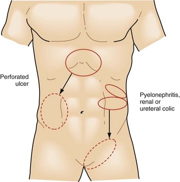
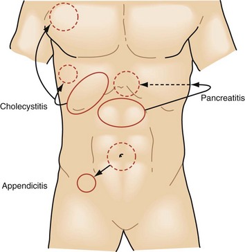
The intensity and severity of the pain are related to the underlying tissue damage. Sudden onset of excruciating pain suggests conditions such as intestinal perforation or arterial embolization with ischemia, although other conditions, such as biliary colic, can present suddenly as well. Pain that develops and worsens over several hours is typical of conditions of progressive inflammation or infection such as cholecystitis, colitis, and bowel obstruction. The history of progressive worsening versus intermittent episodes of pain can help differentiate infectious processes that worsen with time compared with the spasmodic colicky pain associated with bowel obstruction, biliary colic from cystic duct obstruction, or genitourinary obstruction (Figs. 47-2 to 47-4).


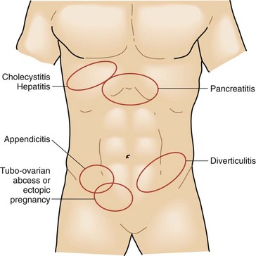
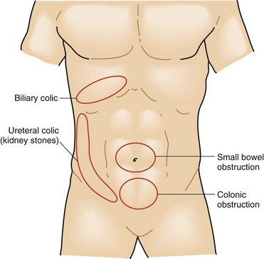
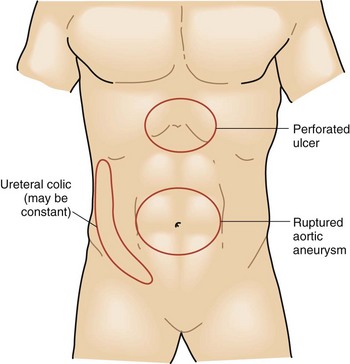
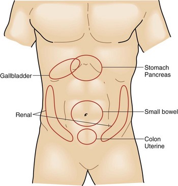
Equally as important as the character of the pain is its location and radiation. Tissue injury or inflammation can trigger visceral and somatic pain. Solid organ visceral pain in the abdomen is generalized in the quadrant of the involved organ, such as liver pain across the right upper quadrant of the abdomen. Small bowel pain is perceived as poorly localized periumbilical pain, whereas colon pain is centered between the umbilicus and pubis symphysis. As inflammation expands to involve the peritoneal surface, parietal nerve fibers from the spine allow for focal and intense sensation. This combination of innervation is responsible for the classic diffuse periumbilical pain of early appendicitis that later shifts to become an intense focal pain in the right lower abdomen at McBurney’s point. If the physician focuses on the character of the current pain and does not thoroughly investigate its onset and progression, he or she will miss these strong historical clues (Figs. 47-5 and 47-6). Pain may also extend well beyond the diseased site. The liver shares some of its innervation with the diaphragm and may create referred pain to the right shoulder from the C3-C5 nerve roots. Genitourinary pain is another source of pain that commonly has a radiating pattern. Symptoms are primarily in the flank region, originating from the splanchnic nerves of T11-L1, but pain often radiates to the scrotum or labia via the hypogastric plexus of S2-S4.


Activities that exacerbate or relieve the pain are also important. Eating will often worsen the pain of bowel obstruction, biliary colic, pancreatitis, diverticulitis, or bowel perforation. Food can provide relief from the pain of nonperforated peptic ulcer disease or gastritis. Clinicians will often recognize that they are evaluating peritonitis while taking the history. Patients with peritoneal inflammation will avoid any activity that stretches or jostles the abdomen. They describe worsening of the pain with any sudden body movement and realize that there is less pain if their knees are flexed. The car ride to the hospital can be agonizing, with the patient feeling every bump along the way.


Associated symptoms can be important diagnostic clues. Nausea, vomiting, constipation, diarrhea, pruritis, melena, hematochezia, and/or hematuria can all be helpful symptoms if present and recognized. Vomiting may occur because of severe abdominal pain of any cause or as a result of mechanical bowel obstruction or ileus. Vomiting is more likely to precede the onset of significant abdominal pain in many medical conditions, whereas the pain of an acute surgical abdomen presents first and stimulates vomiting via medullary efferent fibers that are triggered by visceral afferent pain fibers. Constipation or obstipation can be a result of mechanical obstruction or decreased peristalsis. It may represent the primary problem and require laxatives and prokinetic agents, or merely be a symptom of an underlying condition. A careful history should include whether the patient is continuing to pass any gas or stool from the rectum. A complete obstruction is more likely to be associated with subsequent bowel ischemia or perforation caused by the massive distention that can occur. Diarrhea is associated with several medical causes of acute abdomen, including infectious enteritis, inflammatory bowel disease or parasitic contamination. Bloody diarrhea can be seen in these conditions, as well as in colonic ischemia.


The past medical history could be more helpful than any other single part of the patient’s evaluation. Previous illnesses or diagnoses can greatly increase or decrease the likelihood of certain conditions that would otherwise not be strongly considered. Patients may, for example, report that the current pain is similar to the kidney stone passage that they experienced a decade previously. On the other hand, a prior history of appendectomy, pelvic inflammatory disease, or cholecystectomy can significantly influence the differential diagnosis. During the abdominal examination, all scars on the abdomen should be accounted for by the medical history obtained.


A history of medications and the gynecologic history of female patient are also important. Medications can both create acute abdominal conditions or alternatively mask their symptoms. Although a thorough discussion of the impact of all medications is beyond the scope of this chapter, several common drug classes deserve mention. High-dose narcotic use can interfere with bowel activity and lead to obstipation and obstruction. Narcotics can also contribute to spasm of the sphincter of Oddi and exacerbate biliary or pancreatic pain. They can also suppress pain sensation and alter mental status, which can impair the ability to diagnose the condition accurately. Nonsteroidal anti-inflammatory drugs (NSAIDs) are associated with an increased risk of upper gastrointestinal inflammation and perforation; steroids can block protective gastric mucous production by chief cells and reduce the inflammatory reaction to infection, including advanced peritonitis. As a class, immunosuppressive agents increase a patient’s risk of acquiring various bacterial or viral illnesses and also blunt the inflammatory response, diminishing the pain that is present and the overall physiologic response. Anticoagulants are more prevalent in our emergency patients as the population ages. These drugs may be the cause of gastrointestinal bleeds, retroperitoneal hemorrhages, or rectus sheath hematomas. They can also complicate the preoperative preparation of the patient and be the cause of substantial morbidity if their use goes unrecognized. Finally, recreational drugs can play a role in patients with an acute abdomen. Chronic alcoholism is strongly associated with coagulopathy and portal hypertension from liver impairment. Cocaine and methamphetamine can create an intense vasospastic reaction, which can create life-threatening hypertension and cardiac and intestinal ischemia.


Gynecologic health, specifically the menstrual history, is crucial in the evaluation of lower abdominal pain in a young woman. The likelihood of ectopic pregnancy, pelvic inflammatory disease, mittelschmerz, and/or severe endometriosis are all heavily influenced by the details of the gynecologic history.


Little has changed in the technique or goals of history taking since Dr. Zachary Cope first published his classic paper on the diagnosis of acute abdominal pain in 1921.5 An exception is the application of computers to history taking, which has been extensively studied in Europe.610 Data were collected by physicians on detailed standardized forms during history and physical examinations and entered into computers programmed with a medical database of diseases and their associated signs and symptoms. The computer-generated diagnosis, based on mathematical probabilities, was as much as 20% more accurate than physicians who didn’t use computers to help arrive at a diagnosis. Statistically significant improvement was identified in regard to a timely laparotomy, shortened hospital stay, and reduced need for surgery and hospitalization. However, it should be noted that statistically significant improvements in accuracy and efficiency can be realized without computer assistance if similar standardized forms are used for data collection. This has also been observed in the settings of trauma and critical care.


Physical Examination


An organized and thoughtful physical examination is critical to the development of an accurate differential diagnosis and the subsequent treatment algorithm. Despite newer technologies, including high-resolution computed tomography (CT) scanning, ultrasound, and magnetic resonance imaging (MRI), the physical examination remains a key part of a patient’s evaluation and must not be minimized. Skilled clinicians will be able to develop a narrow and accurate differential diagnosis in most of their patients at the conclusion of the history and physical examination. Laboratory and imaging studies can then be used to confirm the suspicions further, reorder the proposed differential diagnosis or, less commonly, suggest unusual possibilities not yet considered.


The physical examination should always begin with a general inspection of the patient, to be followed by inspection of the abdomen itself. Patients with peritoneal irritation will experience worsened pain with any activity that moves or stretches the peritoneum. These patients will typically lie very still in bed during the evaluation and often maintain flexion of their knees and hips to reduce tension on the anterior abdominal wall. Disease states that cause pain without peritoneal irritation, such as ischemic bowel or ureteral or biliary colic, typically cause patients to shift and fidget in bed continually while trying to find a position that lessens their discomfort (Fig. 47-7). Other important clues such as pallor, cyanosis, and diaphoresis may also be observed during the general inspection.


Abdominal inspection should address the contour of the abdomen, including whether it appears distended or scaphoid or whether a localized mass effect is observed. Special attention should be paid to all scars present and, if surgical in nature, should correlate with the surgical history provided. Fascial hernias may be suspected and can be confirmed during palpation of the abdominal wall. Evidence of erythema or edema of skin may suggest cellulitis of the abdominal wall, whereas ecchymosis is sometimes observed with deeper necrotizing infections of the fascia or abdominal structures, such as the pancreas.


Auscultation can provide useful information about the gastrointestinal tract and vascular system. Bowel sounds are typically evaluated for their quantity and quality. A quiet abdomen suggests an ileus, whereas hyperactive bowel sounds are found in enteritis and early ischemic intestine. The pitch and pattern of the sounds are also considered. Mechanical bowel obstruction is characterized by high-pitched tinkling sounds that tend to come in rushes and are associated with pain. Far away, echoing sounds are often present when significant luminal distention exists. Bruits heard within the abdomen reflect turbulent blood flow in the vascular system. These are most frequently encountered in the setting of high-grade arterial stenoses (70% to 95% but can also be heard if an arteriovenous fistula is present). The clinician can also perform a subtle test for the location and degree of pain during the auscultatory examination by varying the position and amount of pressure applied with the stethoscope. These data can then be compared with the findings during palpation and evaluated for consistency. Even though few patients will try to deceive their physician intentionally, some may exaggerate their pain complaints so as not to be disregarded or taken lightly.


Percussion is used to assess for gaseous distention of the bowel, free intra-abdominal air, degree of ascites, and/or presence of peritoneal inflammation. Hyperresonance, commonly termed tympany to percussion, is characteristic of underlying gas-filled loops of bowel. In the setting of bowel obstruction or ileus, this tympany is heard throughout all but the right upper quadrant, where the liver lies beneath the abdominal wall. If localized dullness to percussion is identified anywhere other than the right upper quadrant, an abdominal mass displacing the bowel should be considered. When liver dullness is lost and resonance is uniform throughout, free intra-abdominal air should be suspected. This air rises and collects beneath the anterior abdominal wall when the patient is in a supine position. Ascites is detected by looking for fluctuance of the abdominal cavity. A fluid wave or ripple can be generated by a quick firm compression of the lateral abdomen. The resulting wave should then travel across the abdominal wall. Movement of adipose tissue in the obese abdomen can be mistaken for a fluid wave. False-positive examinations can be reduced by first pressing the ulnar surface of the examiner’s open palm into the midline soft tissue of the abdominal wall to minimize any movement of the fatty tissue while generating the wave with the opposite hand.


Peritonitis is also assessed by percussion. Older, traditional writings have presented a technique of deep compression of the abdominal wall, followed by abrupt release. This practice is excruciating in the setting of peritoneal inflammation and can create significant discomfort, even in its absence. More sensitive and reliable methods can and should be used. Firmly tapping the iliac crest, flank, or heel of an extended leg will jar the abdominal viscera and elicit characteristic pain when peritonitis is present.


The final major step in the abdominal examination is palpation. Palpation typically provides more information than any other component of the abdominal examination. In addition to revealing the severity and exact location of the abdominal pain, palpation can further confirm the presence of peritonitis and identify organomegaly or an abnormal mass lesion. Palpation should always begin gently and away from the reported area of pain. If considerable pain is induced at the outset of palpation, the patient is likely to guard voluntarily and will continue to do so, limiting the information obtained. Involuntary guarding, or abdominal wall muscle spasm, is a sign of peritonitis and must be distinguished from voluntary guarding. To accomplish this, the examiner applies consistent pressure to the abdominal wall, away from the point of maximal pain, while asking the patient to take a slow deep breath. In the setting of voluntary guarding, the abdominal muscles will relax during the act of inspiration; if involuntary, they remain spastic and tense.


Pain, when focal, suggests an early or well-localized disease process, whereas diffuse pain on palpation is present with extensive inflammation or a late presentation. If pain is diffuse, careful investigation should be carried out to determine where the pain is greatest. Even in the setting of extreme contamination from perforated peptic ulcers or colonic diverticula, the site of maximal tenderness often indicates the underlying source.


Numerous unique physical findings have come to be associated with specific disease conditions and are well described as examination signs (Table 47-1). Murphy’s sign of acute cholecystitis results when inspiration during palpation of the right upper quadrant results in sudden worsening of pain because of descent of the liver and gallbladder toward the examiner’s hand. Several signs help localize the site of underlying peritonitis, including obturator, psoas, and Rovsing’s signs. Others, such as the Fothergill and Carnett signs, help distinguish intra-abdominal disease from that of the abdominal wall.


Table 47-1 Abdominal Examination Signs


SIGN DESCRIPTION DIAGNOSIS OR CONDITION
Aaron Pain or pressure in epigastrium or anterior chest with persistent firm pressure applied to McBurney’s point Acute appendicitis
Bassler Sharp pain created by compressing appendix between abdominal wall and iliacus Chronic appendicitis
Blumberg Transient abdominal wall rebound tenderness Peritoneal inflammation
Carnett Loss of abdominal tenderness when abdominal wall muscles are contracted Intra-abdominal source of abdominal pain
Chandelier Extreme lower abdominal and pelvic pain with movement of cervix Pelvic inflammatory disease
Charcot Intermittent right upper abdominal pain, jaundice, and fever Choledocholithiasis
Claybrook Accentuation of breath and cardiac sounds through abdominal wall Ruptured abdominal viscus
Courvoisier Palpable gallbladder in presence of jaundice Periampullary tumor
Cruveihier Varicose veins at umbilicus (caput medusa) Portal hypertension
Cullen Periumbilical bruising Hemoperitoneum
Danforth Shoulder pain on inspiration Hemoperitoneum
Fothergill Abdominal wall mass that does not cross midline and remains palpable when rectus contracted Rectus muscle hematomas
Grey Turner Local areas of discoloration around umbilicus and flanks Acute hemorrhagic pancreatitis
Iliopsoas Elevation and extension of leg against resistance creates pain Apppendicitis with retrocecal abscess
Kehr Left shoulder pain when supine and pressure placed on left upper abdomen Hemoperitoneum (especially from splenic origin)
Mannkopf Increased pulse when painful abdomen palpated Absent if malingering
Murphy Pain caused by inspiration while applying pressure to right upper abdomen Acute cholecystitis
Obturator Flexion and external rotation of right thigh while supine creates hypogastric pain Pelvic abscess or inflammatory mass in pelvis
Ransohoff Yellow discoloration of umbilical region Ruptured common bile duct
Rovsing Pain at McBurney’s point when compressing the left lower abdomen Acute appendicitis
Ten Horn Pain caused by gentle traction of right testicle Acute appendicitis

A digital rectal examination needs to be performed in all patients with acute abdominal pain, checking for the presence of a mass, pelvic pain, or intraluminal blood. A pelvic examination should be included for all women when evaluating pain located below the umbilicus. Gynecologic and adnexal processes are best characterized by a thorough speculum and bimanual evaluation.

< div class='tao-gold-member'>

Stay updated, free articles. Join our Telegram channel

Aug 1, 2016 | Posted by in CARDIAC SURGERY | Comments Off on Acute Abdomen

Full access? Get Clinical Tree

Get Clinical Tree app for offline access