Acquired Heart Disease: Valvular

Chapter 61 Acquired Heart Disease


Valvular


Stenotic or regurgitant cardiac valves create hemodynamic demands on one or both ventricles of the heart. The compensatory mechanisms of the ventricles permit the heart to tolerate these lesions for varying periods of time, sometimes years, before surgical intervention is required. Ultimately, however, significant valvular lesions produce systolic and/or diastolic ventricular dysfunction, leading to heart failure. As a general rule, surgery for stenotic valve lesions may be deferred until the patient develops symptoms. Regurgitant valve lesions, however, may produce significant ventricular dysfunction before symptoms develop; therefore, surgery in patients who do not have symptoms may be indicated. Among the heart’s valves, the aortic and mitral valves are the most likely to acquire disease; therefore, this chapter focuses on diseases of the aortic and mitral valves.


Historical Perspective


Heart failure from mitral stenosis was well recognized by the late 19th century, and efforts at surgical correction began well before the heart-lung machine was available.1 In 1897, Samways suggested (but never acted on) the possibility of dilating the stenotic mitral valve. Based on his postmortem studies of rheumatic heart disease in London, Brunton in 1902 proposed surgical intervention for mitral stenosis by passing a dilator through the wall of the left ventricle retrograde into the mitral valve orifice. His proposal was shunned by London physicians, and Brunton never tried this maneuver. The concept, however, was applied 20 years later in Boston when the first report of successful surgical correction of mitral stenosis appeared in 1923. Cutler and Levine reported successful relief of mitral stenosis by incision of the valve with a knife introduced through an apical left ventriculotomy. In 1925, Soutter performed the first successful closed mitral commissurotomy at the London Hospital by introducing his index finger through the left atrial appendage. Despite Soutter’s success, he received no more patient referrals, and another 20 years elapsed before the procedure became widespread. In June 1948, Bailey in Philadelphia and Harken in Boston each performed a successful closed mitral commissurotomy. Thereafter, it became widely used to treat mitral stenosis.


By the mid-1970s, the closed technique was supplanted by open mitral commissurotomy. Although closed mitral commissurotomy achieved good palliation of mitral stenosis for its era, open mitral commissurotomy offered several advantages. First, the valvuloplasty could be performed under direct vision. The primary reason for failure of closed mitral commissurotomy was residual stenosis, not restenosis. In up to 75% of patients, the subvalvular apparatus of the mitral valve contributes significantly to the stenosis. The open technique permitted precise and maximal division of fused commissures, as well as fused chordae. In addition, calcium could be sharply débrided from the valve, and any residual mitral insufficiency could be corrected at the time of operation. Finally, the closed technique had the disadvantage of potentially dislodging a left atrial thrombus, resulting in intraoperative embolization and stroke. Today, however, open mitral commissurotomy is rarely performed; it was supplanted by balloon mitral valvuloplasty by the mid-1990s.


Surgical attempts to correct aortic stenosis also began in the early 20th century.1 In 1912, Tuffier, in Paris, unsuccessfully attempted transaortic digital dilation of a stenotic aortic valve. In Charleston, South Carolina, in 1948, Smithy performed the first successful aortic valvotomy in a 21-year-old woman from Ohio. Smithy died later that same year of aortic stenosis at the age of 34 years. Three years later, in Philadelphia, Bailey reported successful aortic valvotomy by insertion of a mechanical dilator across the stenotic valve of patients to open fused commissures. In 1952, Hufnagel and Harvey at Georgetown University placed the first prosthetic ball valve into the descending aorta of a patient with aortic insufficiency. Surgery on the aortic valve under direct vision required the development of cardiopulmonary bypass by Gibbon in 1954. In 1955, Swann performed the first successful aortic valvotomy using hypothermia and inflow occlusion. Initially, open aortic valve operations were limited to aortic valve commissurotomy and débridement of calcified aortic valve leaflets. Harken, in Boston, in 1960, and Starr, in Portland, Oregon, in 1963, however, reported replacement of the aortic valve with a prosthesis. In 1962, Ross in London successfully performed orthotopic homograft valve replacement. In 1967, Ross performed the first pulmonary autograft procedure (Ross procedure) for correction of aortic stenosis. In the mid-1960s, stent-mounted porcine aortic valves were implanted, but these formaldehyde-fixed valves degenerated rapidly. In 1974, Carpentier, in Paris, reported superior longevity of the glutaraldehyde-preserved porcine valve; thereafter, their usage was well established. By 1981, bileaflet mechanical valves were widely implanted in the aortic and mitral positions and largely supplanted the use of ball cage mechanical valves. In the mid-1990s, bovine pericardial valves were shown to have durability similar to porcine valves and both types of bioprostheses became widely implanted. By 2004, most valves implanted in the United States were tissue valves. In 2002, transcatheter aortic valve replacement was performed by Cribier in Rouen, France.


Mitral Valve


Surgical Anatomy of the Mitral Valve


The normal function of the mitral valve is dependent on coordinated interaction of the mitral valve apparatus, which includes the mitral valve annulus, valve leaflets, valve chordae tendineae, and left ventricular papillary muscles. The normal mitral valve has two leaflets, the anterior (or aortic) and posterior or (mural) leaflet. Two papillary muscles arise from the left ventricular wall, the posterior (or posteromedial) and anterior (or anterolateral). Each leaflet of the mitral valve is connected to each of the papillary muscles by tendons, the chordae tendineae.


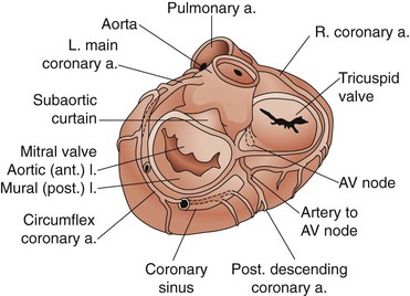
The leaflets are suspended from the mitral annulus, a collagenous structure that encircles the orifice between the left atrium and ventricle. Although the two leaflets have approximately the same surface area, they have different shapes (Fig. 61-1). The anterior leaflet is rectangular. Its base is attached to the mitral annulus anteriorly and the width of the base is approximately one third the circumference of the mitral annulus. This attachment of the anterior leaflet to the mitral annulus extends to the aortic annulus through fibrous tissue, providing fibrous continuity between the aortic and mitral valves; the left ventricular side of the anterior leaflet of the mitral valve is visible immediately as the surgeon looks down through the aortic valve into the left ventricle. The posterior leaflet is rectangular and its attachment to the mitral annulus extends for approximately two thirds of the circumference of the mitral annulus. The two leaflets are separated by two distinct commissures.


There are three important surgical landmarks (see Fig. 61-1). First, the circumflex coronary artery runs along the epicardial surface of the heart overlying the posterior mitral annulus. Only millimeters of left atrial muscle separate the artery from the annulus, making it susceptible to injury during mitral valve surgery. Second, the aortic valve is in close approximation to the anterior leaflet of the mitral valve (aortomitral continuity). The noncoronary leaflet of the aortic valve is therefore susceptible to injury during mitral surgery. Third, the atrioventricular node is located deep to the posteromedial commissure of the mitral valve.


Mitral Stenosis


Causes


Rheumatic fever is the principal cause of mitral stenosis, and approximately two thirds of patients with rheumatic mitral stenosis are female. Rheumatic fever usually occurs in childhood or adolescence (mean age, 8 to 12 years) and creates an inflammatory infiltration of the myocardium and valves. Perhaps because the disease afflicts young people and many years pass before symptoms are manifest, a prior history of rheumatic fever is often difficult to confirm. As the mitral valve heals after acute rheumatic fever, the mitral apparatus may become deformed slowly and the patient typically remains asymptomatic for at least 10 years. Symptoms most commonly appear during the patient’s third or fourth decade of life. Healing of the inflammation from rheumatic fever ultimately causes the cusps and commissures of the mitral valve to thicken and fuse, with concomitant fusion and shortening of the chordae tendineae. The structure of the valve apparatus then calcifies and narrows, becoming funnel-shaped. Such thickening and fusion of the valve not only creates stenosis but also often prevent complete closure of the valve. Of all patients with rheumatic mitral valve disease, approximately 50% have combined mitral stenosis and mitral regurgitation.


Other causes of mitral stenosis that are far less common than rheumatic fever include malignant carcinoid, systemic lupus erythematosus, and rheumatoid arthritis. Rarely, congenital malformation of the valve may cause mitral stenosis, and congenital mitral stenosis is almost never an isolated congenital cardiac lesion.


Pathophysiology


The cross-sectional area of the normal mitral valve is 4 to 6 cm2. A mitral valve area of 2 cm2 is considered moderate mitral stenosis and an area of 1 cm2 is considered severe mitral stenosis. Under normal conditions, there is no pressure gradient across the mitral valve and the left atrial pressure is usually less than 15 mm Hg. As the mitral valve becomes more narrowed, an increasing pressure gradient is required to move the blood across the mitral valve from the left atrium into the left ventricle during diastole; a transvalvular gradient of 10 mm Hg indicates severe mitral stenosis. The significance of the transvalvular gradient is that left atrial pressure progressively increases as the mitral valve becomes more stenotic. In turn, the increased left atrial pressure is transmitted retrograde into the pulmonary veins, pulmonary capillaries, and ultimately pulmonary arteries. A left atrial pressure of approximately 25 mm Hg increases pulmonary capillary pressure enough to produce pulmonary edema.


The severity of obstruction across the valve is determined by the transvalvular gradient and flow rate across the valve. The flow rate is a function of cardiac output and heart rate; because flow across the mitral valve occurs during diastole and diastole is shortened as heart rate increases, a faster heart rate at any given cardiac output increases the transvalvular gradient and raises left atrial pressure. The contribution of the atrial contraction (kick) to cardiac output is particularly important in mitral stenosis; it accomplishes as much as 30% of the transvalvular gradient. Thus, the onset of symptoms is generally associated with exertional activities or loss of the atrial kick with the onset of atrial fibrillation.


To maintain adequate left ventricular filling across a 1-cm2 valve, for example, a pressure gradient of 20 mm Hg is required. A normal left ventricular end-diastolic pressure of 5 mm Hg results in a left atrial pressure of 25 mm Hg. Left atrial pressure rises further if flow rate across the valve increases (increased cardiac output), transit time across the valve is shortened (decreased diastolic time), or atrial kick is lost (atrial fibrillation).


Pulmonary hypertension is an important component of the pathophysiology of mitral stenosis and, when severe, may dominate the clinical picture. At least three pathophysiologic mechanisms contribute to the pulmonary hypertension seen in long-standing mitral valvular disease: (1) increased left atrial pressure transmitted retrograde into the arterial circulation; (2) vascular remodeling of the pulmonary vasculature in response to chronic obstruction to pulmonary venous drainage (fixed component); and (3) pulmonary arterial vasoconstriction (reactive component).


Diagnosis


Diagnostic Tests


Treatment


The symptom-free patient in sinus rhythm requires only prophylaxis against bacterial endocarditis. When symptoms appear, medical treatment of mitral stenosis includes diuretics to lower left atrial pressure and efforts to maintain sinus rhythm with beta-blocking agents or calcium channel blocking agents. Digoxin may be helpful in controlling ventricular rate in patients who do go into atrial fibrillation. Patients in atrial fibrillation may require chronic anticoagulation with warfarin (Coumadin) therapy to lower the risk for systemic embolization.


Mechanical intervention for mitral stenosis should be considered when patients develop symptoms, evidence of pulmonary hypertension appears, or the mitral valve area is reduced to approximately 1 cm2. Other conditions that should prompt surgical consideration include systemic embolization, worsening pulmonary hypertension, and endocarditis. The options for mechanical intervention for mitral stenosis include balloon mitral valvuloplasty, open surgical mitral valvuloplasty (commissurotomy), and mitral valve replacement.


Balloon Mitral Valvuloplasty


First performed in 1984, balloon mitral valvuloplasty has become the treatment of choice for select patients with mitral stenosis.4 Echocardiography may be used to determine patients considered to be good candidates, including those with pliable valve leaflets but without significant valvular calcification or deformation of the chordae tendineae. Contraindications to this procedure include the presence of moderate mitral regurgitation, thickening and calcification of the mitral leaflets, and scarring and calcification of the subvalvular apparatus.5 Performed in the cardiac catheterization suite under fluoroscopic guidance, the technique entails advancement of one or two balloon catheters across the interatrial septum and inflation of the balloon within the stenotic mitral valve.


Balloon mitral valvuloplasty has provided good short-term and intermediate-term results in appropriately selected patients. Balloon inflation should increase the mitral valve area to approximately 2 cm2. This increase in mitral valve area is usually associated with a significant decline in left atrial pressure and transvalvular gradient and with at least a 20% increase in cardiac output. The mortality rate associated with balloon mitral valvuloplasty is 0.5% to 2%. Other risks associated with this procedure include systemic embolism, cardiac perforation, and creation of mitral regurgitation; the risk of each of these complications is approximately 1% to 2%. Increased pulmonary vascular resistance has been shown to decline after successful balloon valvuloplasty. Approximately 10% of patients are left with a residual interatrial septal defect. Three years after balloon valvuloplasty, at least 66% of patients are free of subsequent intervention. In appropriately selected patients, the results of balloon valvuloplasty compare favorably with those of surgical valvuloplasty.6


Mitral Valve Replacement


The mitral valve should be replaced when valvuloplasty is precluded by dense calcification of the leaflets or subvalvular apparatus or because of concomitant mitral regurgitation. Regardless of whether a tissue or mechanical prosthesis is implanted, efforts should be made to preserve the continuity between the left ventricular apex and mitral annulus provided by the chordae tendineae. This may be accomplished by preservation of the chordae tendineae at the time of mitral valve replacement.


The contribution of the mitral apparatus to overall left ventricular function is now well appreciated.7 A mechanical advantage is afforded the left ventricle by the connection of its apex (via the papillary muscles) to the mitral annulus through the chordae tendineae; elimination of this connection by removal of the entire mitral apparatus leads to loss of left ventricular function. Convincing data have demonstrated that preservation of at least some of the chordae tendineae at the time of mitral valve replacement results in better long-term left ventricular function than mitral valve replacement with chordal separation. Therefore, if mitral valve replacement is required, efforts should be made to preserve the posterior and, in some cases, anterior leaflets of the native mitral valve.


The operative mortality rate associated with mitral valve replacement for mitral stenosis is 2% to 10%.8 Operative mortality is increased with advanced age and the presence of coronary disease. Pulmonary hypertension typically resolves after valve replacement, but several weeks or months may be required. The 5-year survival rate after replacement is 70% to 90%.6,9


Mitral Regurgitation


Causes


Competency of the mitral valve requires an intact mitral valve apparatus. Abnormalities of any component of the mitral valve apparatus may produce mitral regurgitation—the mitral leaflets, chordae tendineae, mitral valve annulus, or papillary muscles. Worldwide, rheumatic fever remains the most common cause of mitral regurgitation; it results in deformity and retraction of the leaflets and shortening of the chordae. The leaflets may be perforated by trauma or infective endocarditis. Calcification of the mitral annulus may result in annular rigidity and prevent valve closure, and mitral annular dilation resultant to left ventricular dilation may similarly preclude leaflet apposition during systole.


Chordal rupture may result from trauma, endocarditis, rheumatic fever, or diseases of collagen formation; chordae to the posterior leaflet rupture more frequently than those to the anterior leaflet. Mitral valve prolapse is found in approximately 2% of the U.S. population and up to 5% of patients with mitral valve prolapse develop mitral regurgitation secondary to chordal elongation or rupture. Coronary artery disease may cause infarction of the papillary muscle, resulting in mitral regurgitation. Infarction in the distribution of the anterior descending coronary artery may necrose the anterolateral papillary muscle, whereas the posteromedial muscle may infarct if blood flow through the posterior descending coronary artery is interrupted. Mitral regurgitation caused by myocardial infarction typically presents as a new murmur several days after infarction.


Pathophysiology


The regurgitant mitral valve offers an alternative route whereby blood may exit the left ventricle. During isovolumetric contraction and systole, blood is preferentially ejected into the low-pressure left atrium. The volume of the regurgitant flow (regurgitant fraction) is dependent on the size of the regurgitant orifice and pressure gradient between the left ventricle and left atrium.


Increased left ventricular afterload or decreased forward left ventricular stroke volume increases left ventricular pressure and thereby increases the pressure gradient between the left ventricle and atrium. The mitral valve annulus is enlarged by dilation of the left ventricle. Therefore, the size of the regurgitant orifice is increased by diminished left ventricular contractility, increased left ventricular preload, and increased afterload. Because the valve leaks during systole, the volume of regurgitant flow also increases as heart rate (number of systoles per minute) increases.


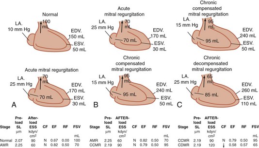
The compensatory mechanism whereby the left ventricle adapts to maintain an adequate systemic blood flow (forward cardiac output) is volume overload; it must pump the combined volume of systemic and regurgitant flows (Fig. 61-2). Volume overload leads to cardiac dilation as well as left ventricular hypertrophy. Because the left ventricle ejects into the reduced resistance of the left atrium, parameters of systolic function (ejection fraction) are increased in mitral regurgitation. As with aortic insufficiency, however, the left ventricle ultimately fails with chronic volume overload. Normal parameters of systolic function indicate significant contractile dysfunction of the left ventricle. An ejection fraction less than 40% in the setting of mitral regurgitation indicates significant left ventricular contractile dysfunction.


image

FIGURE 61-2 Pathophysiology and compensation for acute and chronic mitral regurgitation. A, With acute mitral regurgitation, end-diastolic volume (EDV) increases from 150 to 170 mL. Because the left ventricle ejects blood into both the aorta and the left atrium (LA), end-systolic volume (ESV) decreases from 50 to 30 mL. The ejection fraction therefore increases acutely but, because a significant percentage is ejected into the LA, the volume of blood flow into the aorta (forward stroke volume, FSV) decreases from 100 to 70 mL. The regurgitant volume into the LA increases LA pressure. B, Myocardial compensation for chronic mitral regurgitation includes eccentric left ventricular hypertrophy. Left ventricular EDV increases from 170 to 240 mL. The larger ventricle results in an increased total stroke volume as well as FSV. Enlargement of the LA increases in capacitance, which accommodates the regurgitant volume at a lower pressure. The left ventricular ejection fraction is supernormal. C, Ultimately, the heart decompensates, and the contractile force (CF) of the left ventricle declines; the end-systolic volume increases from 50 to 110 mL. FSV declines. The left ventricle dilates, which further compromises the ability of the mitral valve apparatus to close; the regurgitant volume increases. The ejection fraction remains above normal until contractile function declines further. EF, Ejection fraction; ESS, end-systolic stress; RF, regurgitant fraction; SL, sarcomere length.


(From Carabello BA: Mitral regurgitation: Basic pathophysiologic principles. Mod Concepts Cardiovasc Dis 57:53, 1988.)

< div class='tao-gold-member'>

Stay updated, free articles. Join our Telegram channel

Aug 1, 2016 | Posted by in CARDIAC SURGERY | Comments Off on Acquired Heart Disease: Valvular

Full access? Get Clinical Tree

Get Clinical Tree app for offline access